A Case Report with Lymphangiomatosis of the Colon
- Affiliations
-
- 1Department of Internal Medicine, University of Kyunghee College of Medicine, Seoul, Korea. dramc@hanmail.net
- 2Department of Pathology, University of Kyunghee College of Medicine, Seoul, Korea.
- KMID: 1713849
- DOI: http://doi.org/10.3346/jkms.2010.25.1.155
Abstract
- The incidence of lymphangiomas in the gastrointestinal tract is low, particularly in the colon and rectum, and most cases are solitary. Lymphangiomatosis of the colon are encountered infrequently with only one report in the English literature, and polypectomy was performed for the diagnosis in that case report. However, trends in the diagnosis of lymphangiomatosis of colon have been changing since the development of endoscopic ultrasonography (EUS), and this case is the first in that lymphangiomatosis of the colon was diagnosed without invasive procedures. Here we describe the case of 31-yr-old woman with lymphangiomatosis of the colon with numerous polyposis-like appearing lesions diagnosed by endoscopic ultrasonography and a colonoscopy.
Keyword
MeSH Terms
Figure
-

Fig. 1 Air contrast barium enema showed multiple thumbprint-like lesions (arrows), ranging from 7 to 20 mm in diameter, mainly in the ascending colon.
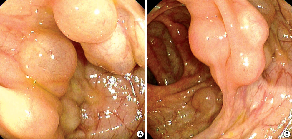
Fig. 2 Endoscopic views of a cluster of elevated lesions, with a smooth surface and gentle slope, in the ascending colon. (A, B) The overlying mucosa was intact and appeared thin, and the lesion was soft and compressible.
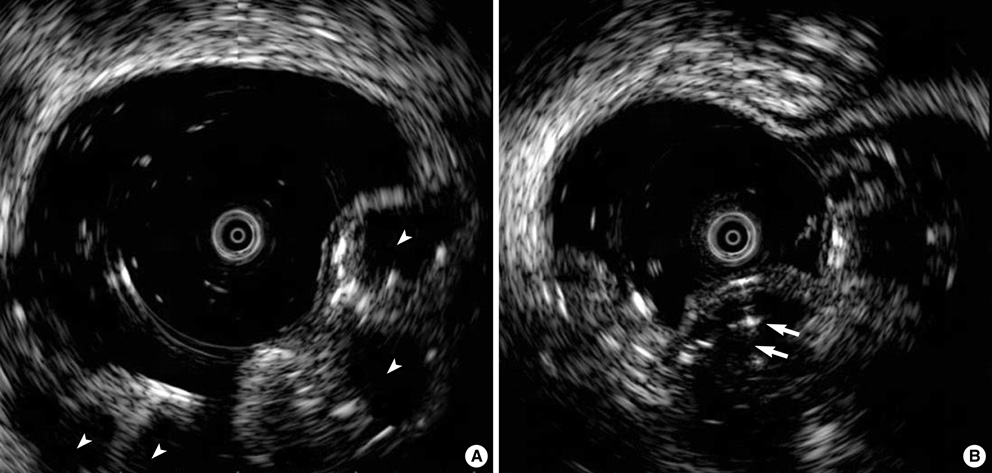
Fig. 3 EUS images of the colon, obtained with a catheter EUS probe (frequency 12 MHz). (A) The EUS image depicting elevated lesions as echo-free cysts (arrowheads) in the submucosal layer. (B) Some submucosal cysts had septal walls (arrows).

Fig. 4 Microscopic findings. (A) Endoscopic biopsy revealed submucosal cyst with occasional multinucleated cells (inset), however, there were no fat or blood cell components (H&E, ×20; inset: H&E, ×400). (B) D2-40 immunostaining showed positive reactivity (arrows) for lining endothelial cells of lymphatic spaces (Polymer method, ×200).
Cited by 1 articles
-
Colonic Lymphangiomatosis Resolved after Excisional Biopsy
Young Soo Lee, Gyu Won Kim, Hye Jae Cho, Chan Sup Shim
Clin Endosc. 2015;48(1):81-84. doi: 10.5946/ce.2015.48.1.81.
Reference
-
1. Watanabe T, Kato K, Sugitani M, Hasunuma O, Sawada T, Hoshino N, Kaneda N, Kawamura F, Arakawa Y, Hirota T. A case of multiple lymphangiomas of the colon suggesting colonic lymphangiomatosis. Gastrointest Endosc. 2000. 52:781–784.
Article2. Yoshitoshi Y, Oda T, Utsumi Y, Kaneko E, Yamashita K. Case of lymphangioma of the ascending colon. Nippon Rinsho. 1976. 23:2264–2267.3. Amaike H, Akioka K, Fujino H, Tanimukai S, Ameno H, Ann T, Nishimoto T, Ikeda E, Muto F, Kurioka H, Hashimoto K, Oouchi T, Tanake K, Harada Y, Ishimine G. A case report of multiple lymphangioma of the colon. Jpn J Surg. 1990. 23:1947–1951.
Article4. Young TH, Ho AS, Tang HS, Hsu CT, Lee HS, Chao YC. Cystic lymphangioma of the transverse colon: report of a case and review of the literature. Abdom Imaging. 1996. 21:415–417.
Article5. Shimizu S, Tada M, Kawai K. Use of endoscopic ultrasonography for the diagnosis of colorectal tumors. Endoscopy. 1990. 22:31–34.
Article6. Kawamoto K, Ueyama T, Iwashita I, Utsunomiya T, Honda H, Onitsuka H, Haraguchi Y, Kojima N, Takano H, Masuda K. Colonic submucosal tumors: comparison of endoscopic US and target air enema CT with barium enema study and colonoscopy. Radiology. 1994. 192:697–702.7. Fujimura Y, Nishishita C, Iida M, Kajihara Y. Lymphangioma of the colon diagnosed with an endoscopic ultrasound probe and dynamic CT. Gastrointest Endosc. 1995. 41:252–254.
Article8. Kochman M, Wiersema M, Hawes R, Canal D, Wiersema L. Preoperative diagnosis of cystic lymphangioma of the colon by endoscopic ultrasound. Gastrointest Endosc. 1997. 45:204–206.
Article9. Hizawa K, Aoyagi K, Kurahara K, Suekane H, Kuwano Y, Nakamura S, Fujishima M. Gastrointestinal lymphangioma: endosonographic demonstration and endoscopic removal. Gastrointest Endosc. 1996. 43:620–624.
Article10. Irisawa A, Bhutani MS. Cystic lymphangioma of the colon: endosonographic diagnosis with through-the-scope catheter miniprobe and determination of further management. Report of a case. Dis Colon Rectum. 2001. 44:1040–1042.11. Kuroda Y, Katoh H, Ohsato K. Cystic lymphangioma of the colon: report of a case and review of the literature. Dis Colon Rectum. 1984. 27:679–682.12. Kuramoto S, Sakai S, Tsuda K, Kaminishi M, Ihara O, Oohara T, Jinbo S, Murakami T. Lymphangioma of the large intestine. Report of a case. Dis Colon Rectum. 1988. 31:900–905.
- Full Text Links
-
- Actions
-
Cited
- CITED
-
- Close
- Share
-
- Similar articles
-
- Colonic Lymphangiomatosis with Normal Colonoscopic Finding in an Adult
- Colonic Lymphangiomatosis Resolved after Excisional Biopsy
- Lymphangiomatosis of Bone and Soft Tissue: A Case Report
- Generalized Lymphangiomatosis: A Case Report
- A Case of Disseminated Lymphangiomatosis Involving Mediastinum, Bone, Spleen and Retroperitoneum in an Asymptomatic Healthy Child
