Current Roles and Future Applications of Cardiac CT: Risk Stratification of Coronary Artery Disease
- Affiliations
-
- 1Department of Cardiology, Cardiovascular Center, Seoul National University Bundang Hospital, Seongnam 463-707, Korea.
- 2Department of Radiology and Research Institute of Radiology, University of Ulsan College of Medicine, Asan Medical Center, Seoul 138-736, Korea. thlim@amc.seoul.kr
- KMID: 1711471
- DOI: http://doi.org/10.3348/kjr.2014.15.1.4
Abstract
- Cardiac computed tomography (CT) has emerged as a noninvasive modality for the assessment of coronary artery disease (CAD), and has been rapidly integrated into clinical cares. CT has changed the traditional risk stratification based on clinical risk to image-based identification of patient risk. Cardiac CT, including coronary artery calcium score and coronary CT angiography, can provide prognostic information and is expected to improve risk stratification of CAD. Currently used conventional cardiac CT, provides accurate anatomic information but not functional significance of CAD, and it may not be sufficient to guide treatments such as revascularization. Recently, myocardial CT perfusion imaging, intracoronary luminal attenuation gradient, and CT-derived computed fractional flow reserve were developed to combine anatomical and functional data. Although at present, the diagnostic and prognostic value of these novel technologies needs to be evaluated further, it is expected that all-in-one cardiac CT can guide treatment and improve patient outcomes in the near future.
MeSH Terms
Figure
-
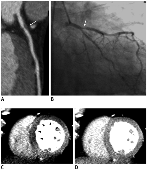
Fig. 1 Case of computed tomography (CT) myocardial perfusion imaging. 54-year-old male patient with chest pain. Coronary CT angiography (A) showed stenosis in proximal left anterior descending artery (white arrow). Invasive coronary angiography (B) also showed presence of significant stenosis in proximal LAD (white arrow), and fractional flow reserve of 0.79 confirmed functional significance of lesion. Stress (C) and rest (D) CT myocardial perfusion imaging showed ischemia (black arrowheads) of LAD territory (images were provided by Dr. Dong Hyun Yang from Asan Medical Center, Korea). LAD = left anterior descending artery.
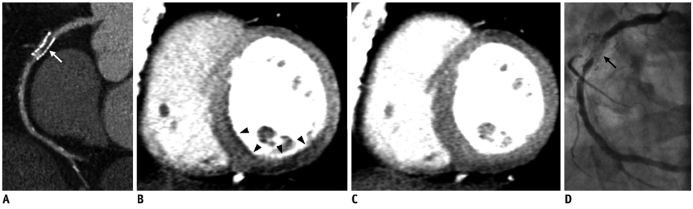
Fig. 2 Case of computed tomography (CT) myocardial perfusion imaging. 64-year-old male patient with history of percutaneous coronary intervention to right coronary artery (RCA) was referred with effort angina. Coronary CT angiography (A) showed stenosis in RCA stent (white arrow), and stress (B) and rest (C) CT myocardial perfusion imaging showed ischemia (black arrowheads) of RCA territory. Invasive coronary angiography (D) confirmed presence of significant in-stent restenosis (black arrow) (Images were provided by Dr. Dong Hyun Yang from Asan Medical Center, Korea).
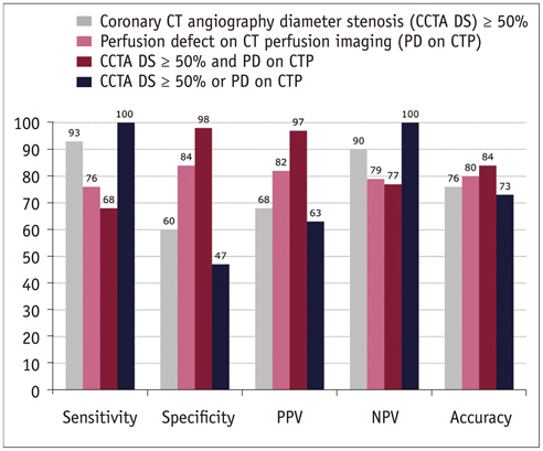
Fig. 3 Diagnostic accuracy of coronary computed tomography (CT) angiography and CT myocardial perfusion imaging to detect functionally significant stenosis, defined as fractional flow reserve ≤ 0.80 (27). PPV = positive predictive value, NPV = negative predictive value
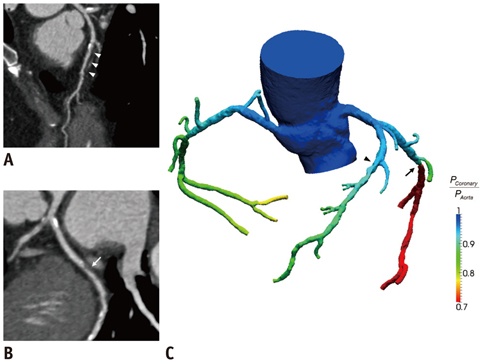
Fig. 4 Case of computed tomography-derived computed fractional flow reserve (FFRCT). 59-year-old female patient complained effort angina. Coronary CT angiography detected (A) diffuse intermediate stenosis of left anterior descending artery (LAD) (white arrowheads) and (B) focal intermediate stenosis of left circumflex artery (LCX) (white arrow). Computation of FFRCT demonstrated that (C) LAD stenosis was not hemodynamically significant with FFRCT value over 0.8 (black arrowhead), and LCX stenosis is ischemia-causing lesion with FFRCT value of less than 0.8 (black arrow) (Images were provided by Dr. Bon-Kwon Koo from Seoul National University Hospital, Korea).
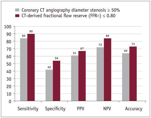
Fig. 5 Diagnostic accuracy of coronary computed tomography (CT) angiography and FFRCT to detect functionally significant stenosis, defined as fractional flow reserve ≤ 0.80 (38). PPV = positive predictive value, NPV = negative predictive value
Cited by 1 articles
-
Diagnostic Accuracy of Electrocardiogram-Gated Thoracic Computed Tomography Angiography without Heart Rate Control for Detection of Significant Coronary Artery Stenosis in Patients with Acute Ischemic Stroke: A Comparative Study
Inyoung Song, Ji Hun Kang, Mi Young Kim, Hweung Kon Hwang, Han Young Kim, Sung Min Ko
Korean J Radiol. 2018;19(5):905-915. doi: 10.3348/kjr.2018.19.5.905.
Reference
-
1. Raggi P, Gongora MC, Gopal A, Callister TQ, Budoff M, Shaw LJ. Coronary artery calcium to predict all-cause mortality in elderly men and women. J Am Coll Cardiol. 2008; 52:17–23.2. Budoff MJ, Shaw LJ, Liu ST, Weinstein SR, Mosler TP, Tseng PH, et al. Long-term prognosis associated with coronary calcification: observations from a registry of 25,253 patients. J Am Coll Cardiol. 2007; 49:1860–1870.3. Greenland P, LaBree L, Azen SP, Doherty TM, Detrano RC. Coronary artery calcium score combined with Framingham score for risk prediction in asymptomatic individuals. JAMA. 2004; 291:210–215.4. Detrano R, Guerci AD, Carr JJ, Bild DE, Burke G, Folsom AR, et al. Coronary calcium as a predictor of coronary events in four racial or ethnic groups. N Engl J Med. 2008; 358:1336–1345.5. Polonsky TS, McClelland RL, Jorgensen NW, Bild DE, Burke GL, Guerci AD, et al. Coronary artery calcium score and risk classification for coronary heart disease prediction. JAMA. 2010; 303:1610–1616.6. Henneman MM, Schuijf JD, Pundziute G, van Werkhoven JM, van der Wall EE, Jukema JW, et al. Noninvasive evaluation with multislice computed tomography in suspected acute coronary syndrome: plaque morphology on multislice computed tomography versus coronary calcium score. J Am Coll Cardiol. 2008; 52:216–222.7. Yoon YE, Chang SA, Choi SI, Chun EJ, Cho YS, Youn TJ, et al. The absence of coronary artery calcification does not rule out the presence of significant coronary artery disease in Asian patients with acute chest pain. Int J Cardiovasc Imaging. 2012; 28:389–398.8. Budoff MJ, Dowe D, Jollis JG, Gitter M, Sutherland J, Halamert E, et al. Diagnostic performance of 64-multidetector row coronary computed tomographic angiography for evaluation of coronary artery stenosis in individuals without known coronary artery disease: results from the prospective multicenter ACCURACY (Assessment by Coronary Computed Tomographic Angiography of Individuals Undergoing Invasive Coronary Angiography) trial. J Am Coll Cardiol. 2008; 52:1724–1732.9. Meijboom WB, Meijs MF, Schuijf JD, Cramer MJ, Mollet NR, van Mieghem CA, et al. Diagnostic accuracy of 64-slice computed tomography coronary angiography: a prospective, multicenter, multivendor study. J Am Coll Cardiol. 2008; 52:2135–2144.10. Miller JM, Rochitte CE, Dewey M, Arbab-Zadeh A, Niinuma H, Gottlieb I, et al. Diagnostic performance of coronary angiography by 64-row CT. N Engl J Med. 2008; 359:2324–2336.11. Hulten EA, Carbonaro S, Petrillo SP, Mitchell JD, Villines TC. Prognostic value of cardiac computed tomography angiography: a systematic review and meta-analysis. J Am Coll Cardiol. 2011; 57:1237–1247.12. Hadamitzky M, Distler R, Meyer T, Hein F, Kastrati A, Martinoff S, et al. Prognostic value of coronary computed tomographic angiography in comparison with calcium scoring and clinical risk scores. Circ Cardiovasc Imaging. 2011; 4:16–23.13. Chow BJ, Wells GA, Chen L, Yam Y, Galiwango P, Abraham A, et al. Prognostic value of 64-slice cardiac computed tomography severity of coronary artery disease, coronary atherosclerosis, and left ventricular ejection fraction. J Am Coll Cardiol. 2010; 55:1017–1028.14. Chow BJ, Small G, Yam Y, Chen L, Achenbach S, Al-Mallah M, et al. Incremental prognostic value of cardiac computed tomography in coronary artery disease using CONFIRM: COroNary computed tomography angiography evaluation for clinical outcomes: an InteRnational Multicenter registry. Circ Cardiovasc Imaging. 2011; 4:463–472.15. Cho I, Shim J, Chang HJ, Sung JM, Hong Y, Shim H, et al. Prognostic value of multidetector coronary computed tomography angiography in relation to exercise electrocardiogram in patients with suspected coronary artery disease. J Am Coll Cardiol. 2012; 60:2205–2215.16. van Werkhoven JM, Schuijf JD, Gaemperli O, Jukema JW, Boersma E, Wijns W, et al. Prognostic value of multislice computed tomography and gated single-photon emission computed tomography in patients with suspected coronary artery disease. J Am Coll Cardiol. 2009; 53:623–632.17. McEvoy JW, Blaha MJ, Nasir K, Yoon YE, Choi EK, Cho IS, et al. Impact of coronary computed tomographic angiography results on patient and physician behavior in a low-risk population. Arch Intern Med. 2011; 171:1260–1268.18. Silber S, Albertsson P, Avilés FF, Camici PG, Colombo A, Hamm C, et al. [Guidelines for percutaneous coronary interventions]. Ital Heart J Suppl. 2005; 6:427–474.19. Smith SC Jr, Feldman TE, Hirshfeld JW Jr, Jacobs AK, Kern MJ, King SB 3rd, et al. ACC/AHA/SCAI 2005 guideline update for percutaneous coronary intervention: a report of the American College of Cardiology/American Heart Association Task Force on Practice Guidelines (ACC/AHA/SCAI Writing Committee to Update 2001 Guidelines for Percutaneous Coronary Intervention). Circulation. 2006; 113:e166–e286.20. Tonino PA, De Bruyne B, Pijls NH, Siebert U, Ikeno F, van't Veer M, et al. Fractional flow reserve versus angiography for guiding percutaneous coronary intervention. N Engl J Med. 2009; 360:213–224.21. Hachamovitch R, Hayes SW, Friedman JD, Cohen I, Berman DS. Comparison of the short-term survival benefit associated with revascularization compared with medical therapy in patients with no prior coronary artery disease undergoing stress myocardial perfusion single photon emission computed tomography. Circulation. 2003; 107:2900–2907.22. Meijboom WB, Van Mieghem CA, van Pelt N, Weustink A, Pugliese F, Mollet NR, et al. Comprehensive assessment of coronary artery stenoses: computed tomography coronary angiography versus conventional coronary angiography and correlation with fractional flow reserve in patients with stable angina. J Am Coll Cardiol. 2008; 52:636–643.23. Gaemperli O, Schepis T, Valenta I, Koepfli P, Husmann L, Scheffel H, et al. Functionally relevant coronary artery disease: comparison of 64-section CT angiography with myocardial perfusion SPECT. Radiology. 2008; 248:414–423.24. Rispler S, Keidar Z, Ghersin E, Roguin A, Soil A, Dragu R, et al. Integrated single-photon emission computed tomography and computed tomography coronary angiography for the assessment of hemodynamically significant coronary artery lesions. J Am Coll Cardiol. 2007; 49:1059–1067.25. Sampson UK, Dorbala S, Limaye A, Kwong R, Di Carli MF. Diagnostic accuracy of rubidium-82 myocardial perfusion imaging with hybrid positron emission tomography/computed tomography in the detection of coronary artery disease. J Am Coll Cardiol. 2007; 49:1052–1058.26. Pazhenkottil AP, Nkoulou RN, Ghadri JR, Herzog BA, Buechel RR, Küest SM, et al. Prognostic value of cardiac hybrid imaging integrating single-photon emission computed tomography with coronary computed tomography angiography. Eur Heart J. 2011; 32:1465–1471.27. Ko BS, Cameron JD, Meredith IT, Leung M, Antonis PR, Nasis A, et al. Computed tomography stress myocardial perfusion imaging in patients considered for revascularization: a comparison with fractional flow reserve. Eur Heart J. 2012; 33:67–77.28. George RT, Jerosch-Herold M, Silva C, Kitagawa K, Bluemke DA, Lima JA, et al. Quantification of myocardial perfusion using dynamic 64-detector computed tomography. Invest Radiol. 2007; 42:815–822.29. Nakauchi Y, Iwanaga Y, Ikuta S, Kudo M, Kobuke K, Murakami T, et al. Quantitative myocardial perfusion analysis using multi-row detector CT in acute myocardial infarction. Heart. 2012; 98:566–572.30. Steigner ML, Mitsouras D, Whitmore AG, Otero HJ, Wang C, Buckley O, et al. Iodinated contrast opacification gradients in normal coronary arteries imaged with prospectively ECG-gated single heart beat 320-detector row computed tomography. Circ Cardiovasc Imaging. 2010; 3:179–186.31. Chow BJ, Kass M, Gagné O, Chen L, Yam Y, Dick A, et al. Can differences in corrected coronary opacification measured with computed tomography predict resting coronary artery flow? J Am Coll Cardiol. 2011; 57:1280–1288.32. Choi JH, Min JK, Labounty TM, Lin FY, Mendoza DD, Shin DH, et al. Intracoronary transluminal attenuation gradient in coronary CT angiography for determining coronary artery stenosis. JACC Cardiovasc Imaging. 2011; 4:1149–1157.33. Choi JH, Koo BK, Yoon YE, Min JK, Song YB, Hahn JY, et al. Diagnostic performance of intracoronary gradient-based methods by coronary computed tomography angiography for the evaluation of physiologically significant coronary artery stenoses: a validation study with fractional flow reserve. Eur Heart J Cardiovasc Imaging. 2012; 13:1001–1007.34. Yoon YE, Choi JH, Kim JH, Park KW, Doh JH, Kim YJ, et al. Noninvasive diagnosis of ischemia-causing coronary stenosis using CT angiography: diagnostic value of transluminal attenuation gradient and fractional flow reserve computed from coronary CT angiography compared to invasively measured fractional flow reserve. JACC Cardiovasc Imaging. 2012; 5:1088–1096.35. Koo BK, Erglis A, Doh JH, Daniels DV, Jegere S, Kim HS, et al. Diagnosis of ischemia-causing coronary stenoses by noninvasive fractional flow reserve computed from coronary computed tomographic angiograms. Results from the prospective multicenter DISCOVER-FLOW (Diagnosis of Ischemia-Causing Stenoses Obtained Via Noninvasive Fractional Flow Reserve) study. J Am Coll Cardiol. 2011; 58:1989–1997.36. Min JK, Leipsic J, Pencina MJ, Berman DS, Koo BK, van Mieghem C, et al. Diagnostic accuracy of fractional flow reserve from anatomic CT angiography. JAMA. 2012; 308:1237–1245.
- Full Text Links
-
- Actions
-
Cited
- CITED
-
- Close
- Share
-
- Similar articles
-
- Cardiac CT
- Assessment of Prognosis and Risk Stratification in Coronary Artery Disease
- Imaging Findings of Coronary Artery Fistula in Children: A Pictorial Review
- A Single Coronary Artery with the Right Coronary Artery Originating from the Left Anterior Descending Artery Detected by Cardiac CT: A Case Report
- Current Progress of Studies of Coronary CT for Risk Prediction of Major Adverse Cardiovascular Event (MACE)
