A Case of Basaloid Squamous Cell Carcinoma of Rectosigmoid Colon
- Affiliations
-
- 1Department of Internal Medicine, Inje University Sanggye Paik Hospital, Inje University College of Medicine, Seoul, Korea. drjtj@paik.ac.kr
- KMID: 1711295
- DOI: http://doi.org/10.4166/kjg.2013.62.6.375
Abstract
- Basaloid squamous cell carcinoma is a rare and aggressive variant of squamous cell carcinoma, which mostly occurs in the upper aerodigestive tracts. Basaloid squamous cell carcinoma also typically arises in the anal canal, but is extremely rare in the lower gastrointestinal tract. A 70-year-old man presented with loose stool and intermittent hematochezia 2 months ago. Colonoscopy showed an ulceroinfiltrative mass on the rectosigmoid colon from 16 cm to 18 cm above the anal verge. Conventional colonoscope could not pass through the lesion but it was possible with pediatric colonoscope. Abdominal CT scan showed 1.6 cm sized wall thickening with circumferential luminal narrowing in the rectosigmoid colon and multiple ill-defined low density masses in both lobes of the liver. Therefore, colon cancer with liver metastasis was suspected. However, basaloid cells were noted on histologic examination, and they were weakly positive for synaptophysin on immunohistochemical study. After palliative lower anterior resection, histologic examination of the resected specimen revealed basaloid differentiation with keratin pearls, and tumor cells were positively stained with high molecular weighted cytokeratin (34BE12) and CK 5/6. Thus, the patient was finally diagnosed with basaloid squamous cell carcinoma of rectosigmoid colon with distant metastases.
Keyword
MeSH Terms
-
Aged
Carcinoma, Squamous Cell/*diagnosis/pathology/surgery
Colonoscopy
Colorectal Neoplasms/*diagnosis/pathology/surgery
Humans
Immunohistochemistry
Keratins/metabolism
Liver Neoplasms/radiography/secondary
Lung Neoplasms/radionuclide imaging/secondary
Male
Positron-Emission Tomography
Synaptophysin/metabolism
Tomography, X-Ray Computed
Keratins
Synaptophysin
Figure
-
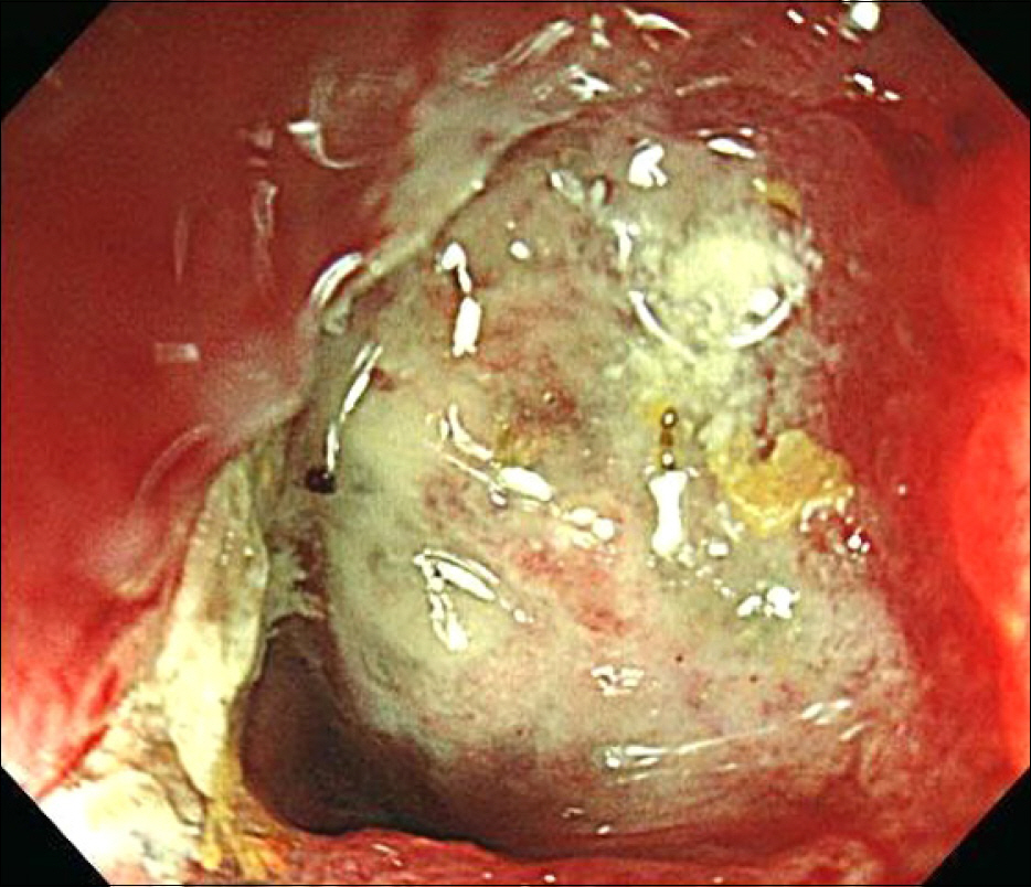
Fig. 1. Colonoscopic finding. A 2 cm sized encircling ulceroinfiltrative mass with luminal narrowing was seen on the rectoscimoid colon from 16 cm to 18 cm above the anal verge.
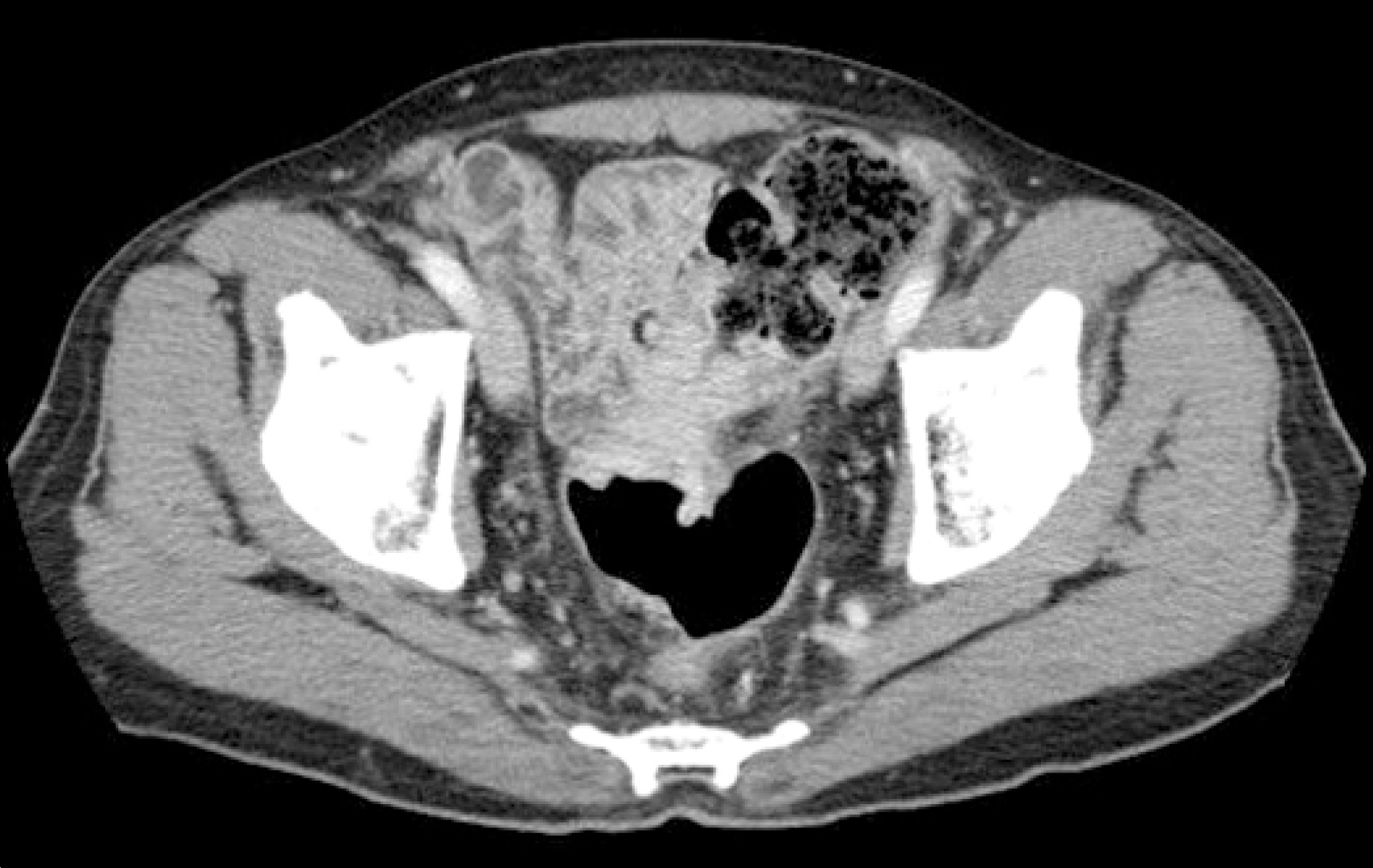
Fig. 2. Abdominal CT finding. A 1.6 cm sized wall thickening with circumferential luminal narrowing was seen on the rectosigmoid junction of colon.
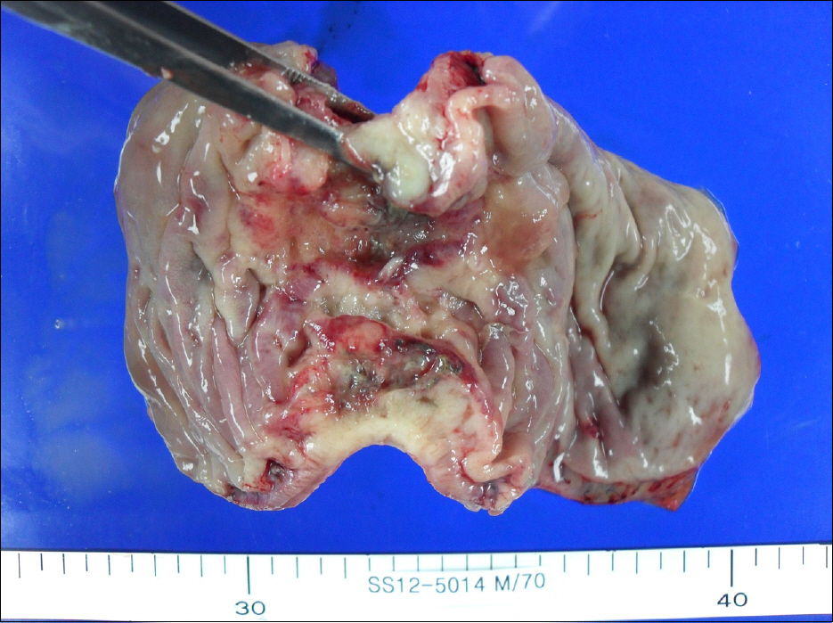
Fig. 3. Gross finding. A 7 cm sized ulceroinfiltrative encircling mass was noted.

Fig. 4. Microscopic findings (H&E). (A) The low power view shows basaloid component and sqaumous component of the tumor (×40). (B) The high power view shows hyperchromatic tumor cells with basaloid differentiation and squamous differentiation with keratin pearls (×400).
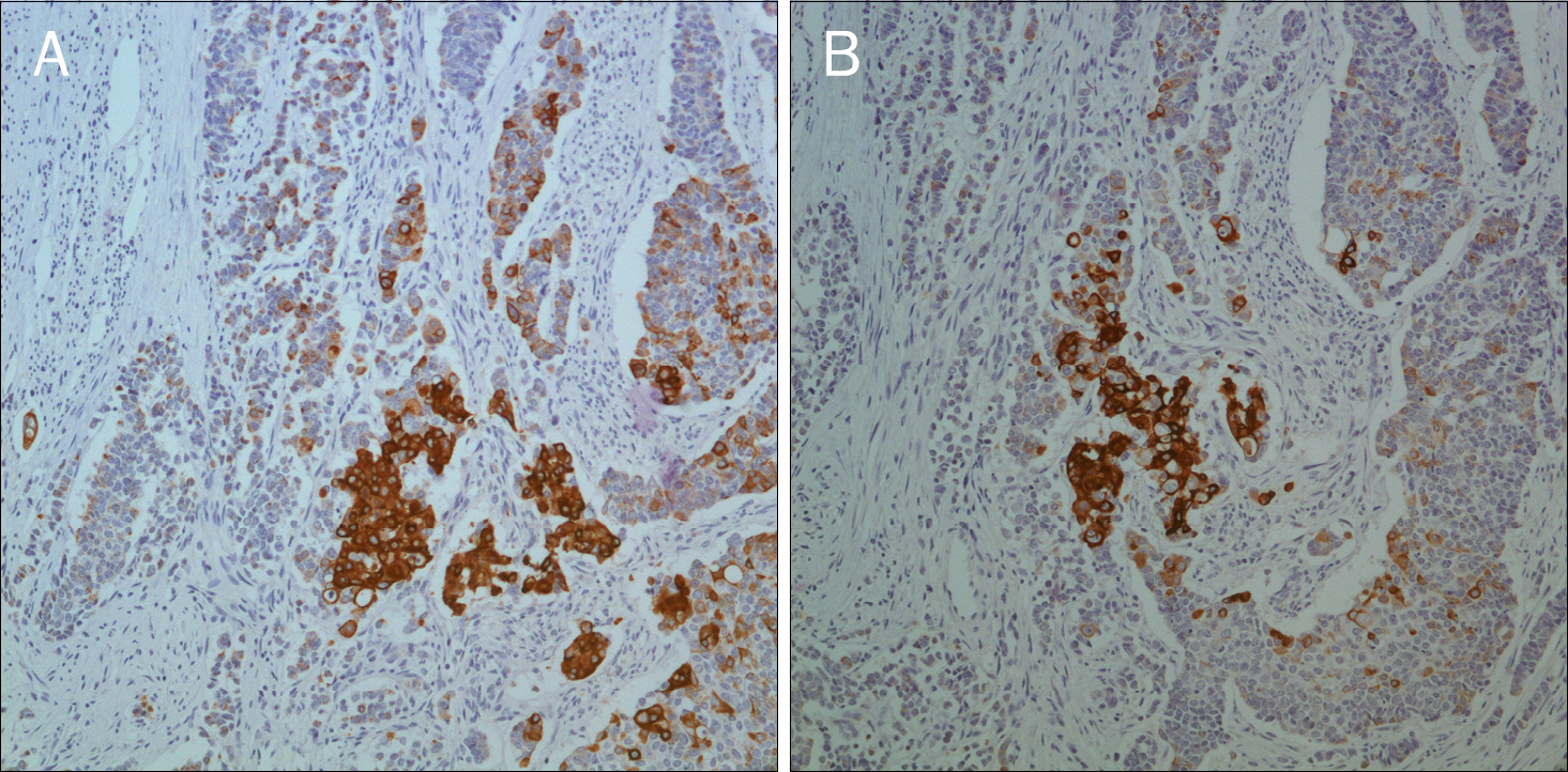
Fig. 5. Immunohistochemical studies.(A) Tumor cells were positive for 34BE12 (×200). (B) Tumor cells were also positive for CK 5/6 (×200).
Reference
-
References
1. Wain SL, Kier R, Vollmer RT, Bossen EH. Basaloid-squamous carcinoma of the tongue, hypopharynx, and larynx: report of 10 cases. Hum Pathol. 1986; 17:1158–1166.2. Newell KJ, Penswick JL, Driman DK. Basaloid carcinoma of the colon arising at the splenic flexure. Histopathology. 2001; 38:232–236.
Article3. Hall-Craggs M, Toker C. Basaloid tumor of the sigmoid colon. Hum Pathol. 1982; 13:497–500.
Article4. Ranaldi R, Sisti S, Librari ML, Suraci V, Bearzi I. Basaloid carcinoma of the sigmoid colon: report of a case. Pathologica. 1988; 80:595–600.5. Jaswal TS, Gupta S, Singh S, Marwah N, Marwah S, Arora B. Basaloid carcinoma of descending colon. Indian J Gastroenterol. 2002; 21:159–160.6. Akbulut S, Cakabay B, Sezgin A, Ozmen CA. Basaloid (cloacogen- ic) carcinoma mimicking intraabdominal abscess: report of a case and review of the literature. Case Rep Gastroenterol. 2009; 3:248–254.7. Park SM, Hur SE, Kwon BJ, et al. Basaloid squamous cell carcinoma of the rectum manifesting as multiple submucosal lesions. Korean J Gastrointest Endosc. 2006; 33:168–172.8. Choussy O, Bertrand M, François A, Blot E, Hamidou H, Dehesdin D. Basaloid squamous cell carcinoma of the head and neck: report of 18 cases. J Laryngol Otol. 2011; 125:608–613.
Article9. Chen SB, Weng HR, Wang G, et al. Basaloid squamous cell carcinoma of the esophagus. J Cancer Res Clin Oncol. 2012; 138:1165–1171.
Article10. Marucci G, Betts CM, Liguori L, Eusebi V. Basaloid carcinoma of the pancreas. Virchows Arch. 2005; 446:322–324.
Article11. Morice WG, Ferreiro JA. Distinction of basaloid squamous cell carcinoma from adenoid cystic and small cell undifferentiated carcinoma by immunohistochemistry. Hum Pathol. 1998; 29:609–612.
Article12. Cho KJ. Basaloid squamous cell carcinoma of the upper aerodigestive tract. Korean J Pathol. 2010; 44:1–8.
Article13. Kim JH, Park JE, Nam JH, et al. A case of synchronous esophageal basaloid squamous carcinoma and cancer of the base of tongue. Korean J Gastrointest Endosc. 2005; 31:383–386.14. Klotz RG Jr, Pamukcoglu T, Souilliard DH. Transitional cloaco-genic carcinoma of the anal canal. Clinicopathologic study of three hundred seventy-three cases. Cancer. 1967; 20:1727–1745.
- Full Text Links
-
- Actions
-
Cited
- CITED
-
- Close
- Share
-
- Similar articles
-
- Basaloid-Squamous Carcinoma of the Esophagus: A case report
- A Case of Basaloid Squamous Cell Carcinoma Occurring in the Mobile Tongue
- Basaloid Squamous Cell Carcinoma of the Lung: Two Case Reports with CT Imaging Findings
- Basaloid-Squamous Cell Carcinoma of the Esophagus: A case report
- A Case of Basaloid Squamous Cell Carcinoma in the Nasal Cavity
