Gyrate Atrophy of the Choroid and Retina Diagnosed by Ornithine-delta-aminotransferase Gene Analysis: A Case Report
- Affiliations
-
- 1Department of Ophthalmology, Samsung Medical Center, Sungkyunkwan University School of Medicine, Seoul, Korea. swkang@skku.edu
- KMID: 1707296
- DOI: http://doi.org/10.3341/kjo.2013.27.5.388
Abstract
- A pair of 19-year-old female identical twins was referred to our hospital with progressive visual loss. They exhibited bilateral chorioretinal atrophy involving the midperiphery on fundoscopy and fluorescein angiography. Bilateral visual field constriction was noted on dynamic Goldmann perimetry, and a markedly impaired response was observed on both photopic and scotopic electroretinograms. Cystoid macular edema was identified in both eyes on optical coherence tomography. Plasma levels of ornithine were elevated. Based on these observations, the patients were diagnosed with gyrate atrophy of the choroid and retina. The clinical diagnosis was confirmed by mutation analysis of the ornithine-delta-aminotransferase (OAT) gene. Patients were treated with a pyridoxine supplement (300 mg/day) and an arginine-restricted diet to lower plasma levels of ornithine, which were successfully reduced without progression of chorioretinal atrophy for 15 months. Our report describes the first case of gyrate atrophy in the Korean population diagnosed by OAT gene analysis and treated with vitamin B6 dietary supplementation.
Keyword
MeSH Terms
-
DNA/*analysis
DNA Mutational Analysis
Diagnosis, Differential
Electroretinography
Female
Fluorescein Angiography
Fundus Oculi
Gyrate Atrophy/*diagnosis/enzymology/genetics
Humans
*Mutation
Ornithine-Oxo-Acid Transaminase/*genetics/metabolism
Tomography, Optical Coherence
Visual Acuity
Young Adult
DNA
Ornithine-Oxo-Acid Transaminase
Figure
-
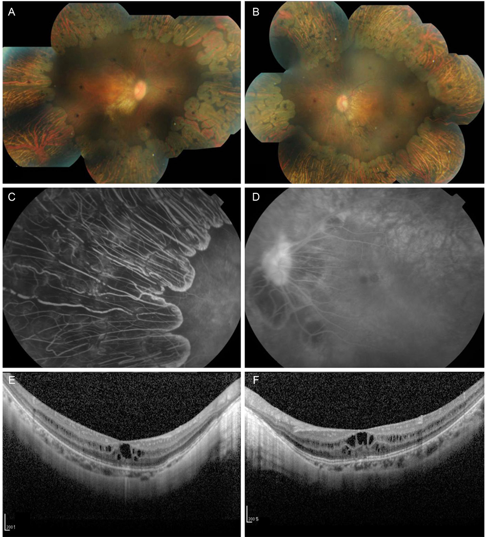
Fig. 1 Panoramic fundus photography, fluorescein angiography, and optical coherence tomography images in both eyes of the older twin. (A,B) The images show coalescent, scalloped chorioretinal atrophy with well-circumscribed, hyperpigmented margins in the midperiphery of the retina and chorioretinal atrophy of the peripapillary area. (C) Fundus fluorescein angiography in the peripheral retina of the right eye exhibiting slight leakage at the margin of chorioretinal atrophy and hyperfluorescence within the atrophic lesion. (D) Fundus fluorescein angiography of the left eye showing minimal leakage and dye accumulation in the macula. (E,F) Optical coherence tomography in both eyes showing a thickened macula simulating cystoid macular edema.
Reference
-
1. Takki KK, Milton RC. The natural history of gyrate atrophy of the choroid and retina. Ophthalmology. 1981; 88:292–301.2. Ramesh V, Shaffer MM, Allaire JM, et al. Investigation of gyrate atrophy using a cDNA clone for human ornithine aminotransferase. DNA. 1986; 5:493–501.3. Inana G, Totsuka S, Redmond M, et al. Molecular cloning of human ornithine aminotransferase mRNA. Proc Natl Acad Sci U S A. 1986; 83:1203–1207.4. Brody LC, Mitchell GA, Obie C, et al. Ornithine delta-aminotransferase mutations in gyrate atrophy. Allelic heterogeneity and functional consequences. J Biol Chem. 1992; 267:3302–3307.5. Simell O, Takki K. Raised plasma-ornithine and gyrate atrophy of the choroid and retina. Lancet. 1973; 1:1031–1033.6. Weleber RG, Kennaway NG. Clinical trial of vitamin B6 for gyrate atrophy of the choroid and retina. Ophthalmology. 1981; 88:316–324.
- Full Text Links
-
- Actions
-
Cited
- CITED
-
- Close
- Share
-
- Similar articles
-
- A Case of Gyrate Atrophy of Choroid and Retina
- Ultra-Structural Changes of the Chorioretina Following Intra-Vitreal Injection of Ornithine in Rabbit
- Choroideremia
- A variant of ornithine aminotransferase from mouse small intestine
- CT and MR Imaging in 3 Patients with Hyperammonemia Due to Ornithine Transcarbamylase Deficiency
