Characteristic Findings of Optical Coherence Tomography in Retinal Angiomatous Proliferation
- Affiliations
-
- 1Myung-Gok Eye Research Institute, Konyang University Kim's Eye Hospital, Seoul, Korea. han66139@kimeye.com
- KMID: 1707289
- DOI: http://doi.org/10.3341/kjo.2013.27.5.351
Abstract
- PURPOSE
To identify the unique pathologic findings of retinal angiomatous proliferation (RAP) in optical coherence tomography (OCT).
METHODS
Retrospectively, 29 eyes of 25 patients with age-related macular degeneration and complicated RAP were analyzed. All 29 eyes had choroidal neovascularization (CNV) in the area of pigment epithelial detachment (PED) or adjacent to it, which was visible with fluorescein angiography or indocyanine green angiography. Cross-sectional images were obtained by OCT scanning through the CNV lesions.
RESULTS
Six distinctive findings of OCT included drusen (100%), inner retinal cyst (80%), outer retinal cyst (68%), fibrovascular PED (84%), serous retinal detachment (40%), and PED (68%).
CONCLUSIONS
Through analysis of OCT findings, we revealed six different types of lesions distinctive of RAP which may provide helpful diagnostic information for subsequent treatment and predicting the prognosis of RAP.
Keyword
MeSH Terms
Figure
-
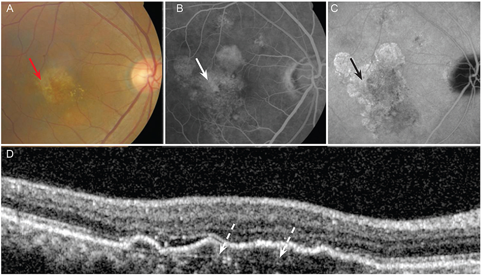
Fig. 1 A 66-year-old man with retinal angiomatous proliferation. (A) Fundus photograph showing drusen, which appears as: confluent yellowish spots on the macula (red arrow). (B) Fluorescein angiogram revealing hyperfluent, ill-defined confluent lesions including focal hyperpigmentation of the macula (arrow). (C) Indocyanine green angiogram showing small drusen, which are mildly hyperfluorescent or not clearly seen (black arrow). (D) Optical coherence tomography scan showing incontiguous elevation of the retinal pigment epithelium (RPE) with slight thickening of RPE/Bruch's membrane complex (dotted arrow).
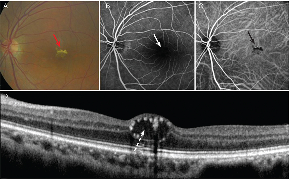
Fig. 2 A 54-year-old man with retinal angiomatous proliferation. (A) Fundus photograph revealing a delicate, star-shaped distortion of the macula without prominent cyst formation (red arrow). (B,C) Fluorescein and indocyanine green angiogram demonstrating retinal distortion, though leakage of dye is not found (arrows). (D) Optical coherence tomography scan showing round, minimally reflective (darker) space spanning nearly the entire thickness of the retina and extending from the retinal pigment epithelium/choriocapillaris reflection to the highly reflective anterior boundary of the neurosensory retina (dotted arrow).
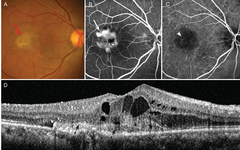
Fig. 3 A 61-year-old woman with retinal angiomatous proliferation. (A) Fundus photograph showing distortion of the macula with yellowish scar change surrounding a cyst-like lesion (red arrow). (B) Fluorescein angiogram revealing dense hyperfluorescence with diffuse leakage of dye in cystic spaces near, but not inside, the fovea (arrow). (C) Indocyanine green angiogram showing the presence of a RAP, which is classified as an irregular hyperfluorescence with termination of the retinal vasculature (arrow head). (D) Optical coherence tomography scan showing subretinal tissue presumably corresponding to the yellowish scar change visible on the color photo (dotted black arrow). Note the inner retinal cystoid cavities above.

Fig. 4 A 65-year-old woman with retinal angiomatous proliferation. (A) Fundus photograph showing diffuse, patch-like cystic change underneath the retinal pigment epithelium in the macula (red arrow). (B) Fluorescein angiogram showing small hyperfluorescence with diffuse leakage of dye in the cystic spaces near and inside the fovea with stippled hyperfluorescence beneath the fovea (arrow). (C) Indocyanine green angiogram revealing the presence of a retinal angiomatous proliferation, which is classified as an irregular hyperfluorescence (black arrow). (D) Optical coherence tomography scan showing two adjacent outer retinal cysts (ORCs) located in a fibrotic area associated with choroidal neovascularization, which is surrounded by a hyperreflective border (dotted arrow). Note the differences between the ORCs and the inner retinal cystoid cavities above.
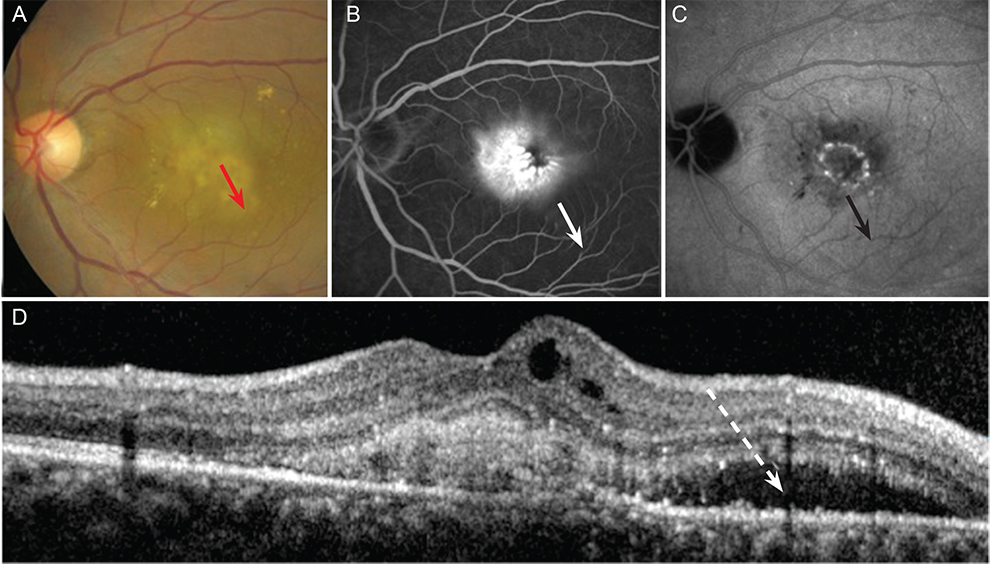
Fig. 5 A 55-year-old woman with retinal angiomatous proliferation. (A) Fundus photograph showing distortion of the macula with yellowish patch-like lesions surrounded by broad cystic change (red arrow). (B) Fluorescein angiogram revealing dense hyperfluorescence with diffuse leakage of dye mimicking choroidal neovascularization in the macula. A large cystic lesion lies inferotemporally, but cannot be clearly seen (arrow). (C) Indocyanine green angiogram showing a more prominent boundary of cystic space below the macula (black arrow). (D) Optical coherence tomography scan indicating subretinal fluid (dotted arrow) adjacent to the subretinal tissue.

Fig. 6 A 64-year-old woman with retinal angiomatous proliferation. (A) Fundus photograph showing a serous retinal pigment epithelium detachment and subretinal hemorrhage (red arrow). (B,C) Fluorescein and indocyanine green angiogram showing dye leakage from choroidal neovascularization within the retinal pigment epithelial detachment (arrow and arrowhead). (D) Optical coherence tomography scan revealing the dome-shaped elevation of a highly reflective layer corresponding to the detached retinal pigment epithelium (dotted arrow).
Reference
-
1. Dell'Omo R, Cassetta M, dell'Omo E, et al. Aqueous humor levels of vascular endothelial growth factor before and after intravitreal bevacizumab in type 3 versus type 1 and 2 neovascularization: a prospective, case-control study. Am J Ophthalmol. 2012; 153:155–161.e2.2. Brancato R, Introini U, Pierro L, et al. Optical coherence tomography (OCT) angiomatous prolifieration (RAP) in retinal. Eur J Ophthalmol. 2002; 12:467–472.3. Rouvas AA, Papakostas TD, Ntouraki A, et al. Angiographic and OCT features of retinal angiomatous proliferation. Eye (Lond). 2010; 24:1633–1642.4. Yannuzzi LA, Negrao S, Iida T, et al. Retinal angiomatous proliferation in age-related macular degeneration. Retina. 2001; 21:416–434.5. Hartnett ME, Weiter JJ, Garsd A, Jalkh AE. Classification of retinal pigment epithelial detachments associated with drusen. Graefes Arch Clin Exp Ophthalmol. 1992; 230:11–19.6. Kuhn D, Meunier I, Soubrane G, Coscas G. Imaging of chorioretinal anastomoses in vascularized retinal pigment epithelium detachments. Arch Ophthalmol. 1995; 113:1392–1398.7. Hartnett ME, Weiter JJ, Staurenghi G, Elsner AE. Deep retinal vascular anomalous complexes in advanced age-related macular degeneration. Ophthalmology. 1996; 103:2042–2053.8. Slakter JS, Yannuzzi LA, Schneider U, et al. Retinal choroidal anastomoses and occult choroidal neovascularization in age-related macular degeneration. Ophthalmology. 2000; 107:742–753.9. Lafaut BA, Aisenbrey S, Vanden Broecke C, Bartz-Schmidt KU. Clinicopathological correlation of deep retinal vascular anomalous complex in age related macular degeneration. Br J Ophthalmol. 2000; 84:1269–1274.10. Yannuzzi LA, Slakter JS, Sorenson JA, et al. Digital indocyanine green videoangiography and choroidal neovascularization. Retina. 1992; 12:191–223.11. Yannuzzi LA, Hope-Ross M, Slakter JS, et al. Analysis of vascularized pigment epithelial detachments using indocyanine green videoangiography. Retina. 1994; 14:99–113.12. Wolf S, Wolf-Schnurrbusch U. Spectral-domain optical coherence tomography use in macular diseases: a review. Ophthalmologica. 2010; 224:333–340.13. Susan BB, Neil M, Bressler . Age-related macular degeneration: non-neovascular early AMD, intermediate AMD, and geographic atrophy. In : Schachat AP, Ryan SJ, editors. Retina. 5th ed. London: Elsevier;2013. p. 1161.14. Gass JD. Serous retinal pigment epithelial detachment with a notch: a sign of occult choroidal neovascularization. Retina. 1984; 4:205–220.15. Wolff B, Maftouhi MQ, Mateo-Montoya A, et al. Outer retinal cysts in age-related macular degeneration. Acta Ophthalmol. 2011; 89:e496–e499.16. Fernandes LH, Freund KB, Yannuzzi LA, et al. The nature of focal areas of hyperfluorescence or hot spots imaged with indocyanine green angiography. Retina. 2002; 22:557–568.17. Zacks DN, Johnson MW. Retinal angiomatous proliferation: optical coherence tomographic confirmation of an intraretinal lesion. Arch Ophthalmol. 2004; 122:932–933.18. Chan WM, Lai TY, Lai RY, et al. Half-dose verteporfin photodynamic therapy for acute central serous chorioretinopathy: one-year results of a randomized controlled trial. Ophthalmology. 2008; 115:1756–1765.19. Khanifar AA, Koreishi AF, Izatt JA, Toth CA. Drusen ultrastructure imaging with spectral domain optical coherence tomography in age-related macular degeneration. Ophthalmology. 2008; 115:1883–1890.20. Cohen SY, Dubois L, Tadayoni R, et al. Prevalence of reticular pseudodrusen in age-related macular degeneration with newly diagnosed choroidal neovascularisation. Br J Ophthalmol. 2007; 91:354–359.21. Leveziel N, Puche N, Richard F, et al. Genotypic influences on severity of exudative age-related macular degeneration. Invest Ophthalmol Vis Sci. 2010; 51:2620–2625.22. Liakopoulos S, Ongchin S, Bansal A, et al. Quantitative optical coherence tomography findings in various subtypes of neovascular age-related macular degeneration. Invest Ophthalmol Vis Sci. 2008; 49:5048–5054.23. Zweifel SA, Engelbert M, Laud K, et al. Outer retinal tubulation: a novel optical coherence tomography finding. Arch Ophthalmol. 2009; 127:1596–1602.24. Doukas J, Mahesh S, Umeda N, et al. Topical administration of a multi-targeted kinase inhibitor suppresses choroidal neovascularization and retinal edema. J Cell Physiol. 2008; 216:29–37.25. Coscas F, Coscas G, Souied E, et al. Optical coherence tomography identification of occult choroidal neovascularization in age-related macular degeneration. Am J Ophthalmol. 2007; 144:592–599.26. Sato T, Iida T, Hagimura N, Kishi S. Correlation of optical coherence tomography with angiography in retinal pigment epithelial detachment associated with age-related macular degeneration. Retina. 2004; 24:910–914.
- Full Text Links
-
- Actions
-
Cited
- CITED
-
- Close
- Share
-
- Similar articles
-
- Comparison of Optical Coherence Tomography Characteristics among Three Subtypes of Exudative Age-related Macular Degeneration
- Optical Coherence Tomography Findings in Best Disease
- Fundus Autofluorescence, Fluorescein Angiography and Spectral Domain Optical Coherence Tomography Findings of Retinal Astrocytic Hamartomas in Tuberous Sclerosis
- Foveal Retinal Detachment Diagnosed by Optical Coherence Tomography after Successful Retinal Detachment Surgery
- Assessment of retinal degeneration with optical coherence tomography in a dog
