Korean J Radiol.
2013 Jun;14(3):497-500. 10.3348/kjr.2013.14.3.497.
Congenital Pial Arteriovenous Fistula in the Temporal Region Draining into Cavernous Sinus: A Case Report
- Affiliations
-
- 1Department of Neurosurgery, West China Hospital of Sichuan University, Chengdu, Sichuan Province, 610041, China. xiaodong_1962@163.com
- 2Department of Neurosurgery, Affiliated Hospital of Hainan Medical College, Haikou, Hainan Province, 570102, China.
- KMID: 1705466
- DOI: http://doi.org/10.3348/kjr.2013.14.3.497
Abstract
- This report concerns a 4-month-old infant with progressive prominent and redness of his left eye since birth. This report concerns a 4-month-old infant with progressive prominent redness of his left eye since birth. Angiography revealed a congenital pial arteriovenous fistula between the temporal branch of the left posterior cerebral artery and left cavernous sinus through the sphenoparietal sinus, a condition not reported in the literature. The fistula was successfully occluded with two micro-coils by vertebrobasilar approach.
MeSH Terms
Figure
-
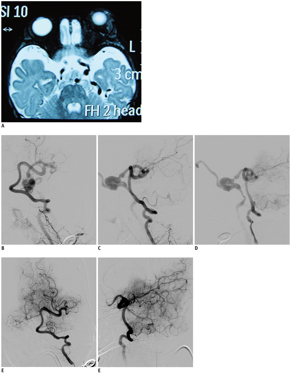
Fig. 1 The MRI/DSA presentation and embolism result of this pial AVF in a 4-month-old infant. A. MRI revealed that proptosis of left eye, prominence of left cavernous sinus and enlargement of extraocular muscles. B, C. DSA revealed pial AVF between temporal branch of left PCA and left cavernous sinus through sphenoparietal sinus (B. AP image, C. LAT image). D. Cerebral angiography was repeated sequentially after coil was filled with checking for occlusion of fistula. E, F. Angiogram of post-embolization revealed fistula was entirely occluded by using only two micro-coils (E. AP image, F. LAT image).
Reference
-
1. Halbach VV, Higashida RT, Hieshima GB, Norman D. Normal perfusion pressure breakthrough occurring during treatment of carotid and vertebral fistulas. AJNR Am J Neuroradiol. 1987. 8:751–756.2. Hoh BL, Putman CM, Budzik RF, Ogilvy CS. Surgical and endovascular flow disconnection of intracranial pial single-channel arteriovenous fistulae. Neurosurgery. 2001. 49:1351–1363. discussion 1363-1364.3. Oka K, Rhoton AL Jr, Barry M, Rodriguez R. Microsurgical anatomy of the superficial veins of the cerebrum. Neurosurgery. 1985. 17:711–748.4. Amaral FT, Machado HR, Almeida S. [Congenital cerebral arteriovenous fistula. Diagnostic peculiarity and surgical repair in an infant with heart failure]. Arq Bras Cardiol. 1994. 63:207–209.5. Haase J. Congestive heart failure secondary to cerebral arteriovenous fistula. Childs Nerv Syst. 1988. 4:75.6. Berant M, Tadmor R, Blieden L, Deutsch V, Neufeld HN. Cerebral arteriovenous fistula causing congestive heart failure in infancy. Angiology. 1977. 28:684–686.7. Sasamori T, Hida K, Yano S, Asano T, Iwasaki Y. Cervical perimedullary arteriovenous fistula in an infant presenting with subarachnoid hemorrhage--case report. Neurol Med Chir (Tokyo). 2008. 48:409–413.8. Lee JY, Son YJ, Kim JE. Intracranial pial arteriovenous fistulas. J Korean Neurosurg Soc. 2008. 44:101–104.9. Nelson K, Nimi Y, Lasjaunias P, Berenstein A. Endovascular embolization of congenital intracranial pial arteriovenous fistulas. Neuroimaging Clin N Am. 1992. 2:309–317.10. Paramasivam S, Toma N, Niimi Y, Berenstein A. De novo development of dural arteriovenous fistula after endovascular embolization of pial arteriovenous fistula. J Neurointerv Surg. 2012. [Epub ahead of print].11. Rivera R, Blanc R, Piotin M, Spelle L, Moret J. Single hole cerebral arteriovenous fistula between the anterior choroidal artery and the basal vein of Rosenthal in a child. Childs Nerv Syst. 2009. 25:1521–1523.
- Full Text Links
-
- Actions
-
Cited
- CITED
-
- Close
- Share
-
- Similar articles
-
- Embolization of Cerebral Pial Arteriovenous Fistula Under Balloon-assisted Flow Control Using NBCA: a Case Report
- Congenital Intracranial Pial Arteriovenous Fistula Complicated with Congestive Heart Failure in Neonate: A Case Report
- Successful Treatment of Intracranial Small Pial Single-Channel Arteriovenous Fistula Using N-butyl Cyanoacrylate: Report of 2 Cases
- Middle temporal vein access for transvenous embolization of Cavernous sinus dural arteriovenous fistula: A case report and review of literature
- Transvenous Embolization of Cavernous Sinus Dural Arteriovenous Fistula Using the Direct Superior Ophthalmic Vein Approach: A Case Report
