Korean J Ophthalmol.
2012 Dec;26(6):428-431. 10.3341/kjo.2012.26.6.428.
Ultrawide-field Fluorescein Angiography for Evaluation of Diabetic Retinopathy
- Affiliations
-
- 1Department of Ophthalmology, Samsung Medical Center, Sungkyunkwan University School of Medicine, Seoul, Korea. oculus@naver.com
- KMID: 1499680
- DOI: http://doi.org/10.3341/kjo.2012.26.6.428
Abstract
- PURPOSE
To investigate the advantages of ultrawide-field fluorescein angiography (FA) over the standard fundus examination in the evaluation of diabetic retinopathy (DR).
METHODS
Ultrawide-field FAs were obtained in 118 eyes of 59 diabetic patients; 11 eyes with no DR, 71 eyes with nonproliferative diabetic retinopathy (NPDR), and 36 eyes with proliferative diabetic retinopathy (PDR), diagnosed by the standard method. The presence of peripheral abnormal lesions beyond the standard seven fields was examined.
RESULTS
Ultrawide-field FA images demonstrated peripheral microaneurysms in six (54.5%) of 11 eyes with no DR and all eyes with moderate to severe NPDR and PDR. Peripheral retinal neovascularizations were detected in three (4.2%) of 71 eyes with NPDR and in 13 (36.1%) of 36 eyes with PDR. Peripheral vascular nonperfusion and vascular leakage were found in two-thirds of eyes with severe NPDR and PDR.
CONCLUSIONS
Ultrawide-field FA demonstrates peripheral lesions beyond standard fields, which can allow early detection and a close evaluation of DR.
MeSH Terms
Figure
-
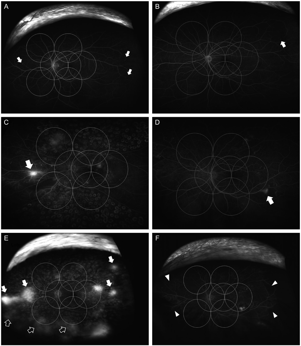
Fig. 1 (A,B) Ultrawide-field fluorescein angiography shows peripheral microaneurysms (arrows) outside the seven standard Early Treatment Diabetic Retinopathy Study (ETDRS) fields in eyes diagnosed as normal fundus with the standard fundus examination. (C,D) Peripheral retinal neovascularizations (arrows) were found outside the standard seven ETDRS fields in eyes diagnosed as nonproliferative diabetic retinopathy by the standard method. (E,F) Multiple lesions of neovascularization (arrows), vascular leakage (blank arrows), and nonperfusion (arrow heads) observed beyond the borders of standard fields in ultrawide-field fluorescein angiography.
Reference
-
1. Furuhashi T, Moroi M, Masai H, et al. Correlation of chronic kidney disease, diabetes and peripheral artery disease with cardiovascular events in patients using stress myocardial perfusion imaging. Ann Nucl Med. 2011. 25:634–642.2. Kaines A, Tsui I, Sarraf D, Schwartz S. The use of ultra wide field fluorescein angiography in evaluation and management of uveitis. Semin Ophthalmol. 2009. 24:19–24.3. Win PH, Young TA. Optos Panoramic200A fluorescein angiography for proliferative diabetic retinopathy with asteroid hyalosis. Semin Ophthalmol. 2007. 22:67–69.4. Manivannan A, Plskova J, Farrow A, et al. Ultra-wide-field fluorescein angiography of the ocular fundus. Am J Ophthalmol. 2005. 140:525–527.5. Anderson L, Friberg TR, Singh J. Ultrawide-angle retinal imaging and retinal detachment. Semin Ophthalmol. 2007. 22:43–47.6. Kaines A, Oliver S, Reddy S, Schwartz SD. Ultrawide angle angiography for the detection and management of diabetic retinopathy. Int Ophthalmol Clin. 2009. 49:53–59.7. Mackenzie PJ, Russell M, Ma PE, et al. Sensitivity and specificity of the optos optomap for detecting peripheral retinal lesions. Retina. 2007. 27:1119–1124.8. Oliver SC, Schwartz SD. Peripheral vessel leakage (PVL): a new angiographic finding in diabetic retinopathy identified with ultra wide-field fluorescein angiography. Semin Ophthalmol. 2010. 25:27–33.
- Full Text Links
-
- Actions
-
Cited
- CITED
-
- Close
- Share
-
- Similar articles
-
- Current Concepts in Diabetic Retinopathy
- The Effect of Fluorescein Angiography on Renal Function in Patients with Diabetic Retinopathy
- New Modalities for the Diagnosis and Treatment of Diabetic Retinopathy
- Clinical Analysis of Diabetic Retinopathy According to the Type of Diabetes Mellitus
- Simplified Correction of Ischemic Index in Diabetic Retinopathy Evaluated by Ultra-widefield Fluorescein Angiography
