Supratentorial Intracerebral Schwannoma : Its Fate and Proper Management
- Affiliations
-
- 1Department of Neurosurgery, Seoul National University Hospital, Seoul, Korea. chungc@snu.ac.kr
- 2Neuroscience Research Institute, Seoul National University Medical Research Center, Seoul, Korea.
- 3Clinical Research Institute, Seoul National University Hospital, Seoul, Korea.
- 4Department of Pathology, Seoul National University Hospital, Seoul, Korea.
- KMID: 1499341
- DOI: http://doi.org/10.3340/jkns.2013.54.4.340
Abstract
- Intracerebral schwannomas are rare and there have been none reported in Korea. We present the case of a 25-year-old man with newly developed right-side weakness and recent seizure aggravation. His seizures started approximately 9 years prior to admission. At that time, a 1 cm diameter intra-axial enhancing mass at the left precentral gyrus was found on magnetic resonance image (MRI). After 9 years of observation and treatment with antiepileptic medication, an MRI taken due to symptom aggravation revealed peri-tumoral cyst formation with tumor enlargement. The tumor was surgically removed. Subsequently, right-side weakness diminished and there was good seizure control. Pathologic diagnosis was schwannoma. Schwannoma is a very rare tumor and there are no pathognomonic findings on radiologic images; thus, it is challenging to make a correct diagnosis. However, considering the natural course and excellent prognosis after surgical treatment of this kind of intra-axial mass with benign features, early surgery for diagnosis and proper treatment is highly recommended.
Figure
-
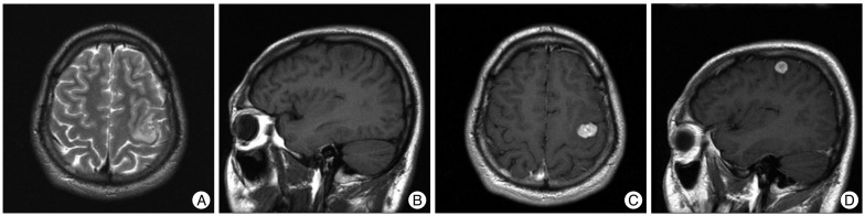
Fig. 1 Brain MRI after the first seizure attack revealed a small mass lesion at the precentral gyrus with peritumoral edema. The mass is of heterogeneously high signal intensity on a T2-weighted axial image (A) and predominantly hypointense to gray matter on a T1-weighted sagittal image (B). It enhances homogeneously after gadolinium injection (C and D).
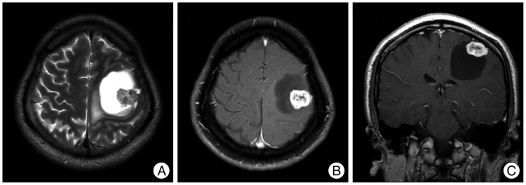
Fig. 2 9-year follow-up brain MRI revealed a slightly increased enhancing mass with a large, newly appeared peritumoral cyst. The cyst is hyperintense on a T2-weighted axial image (A), and hypointense on a T1-weighted axial and coronal images (B and C). Cystic fluid shows a similar signal as the cerebrospinal fiuid.
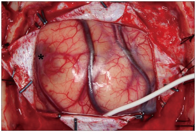
Fig. 3 After opening the dura, a bluish discoloration of the cerebral cortex is observed. Tumor location with the discoloration is marked as '*'. In addition, diffuse swollen brain is observed.
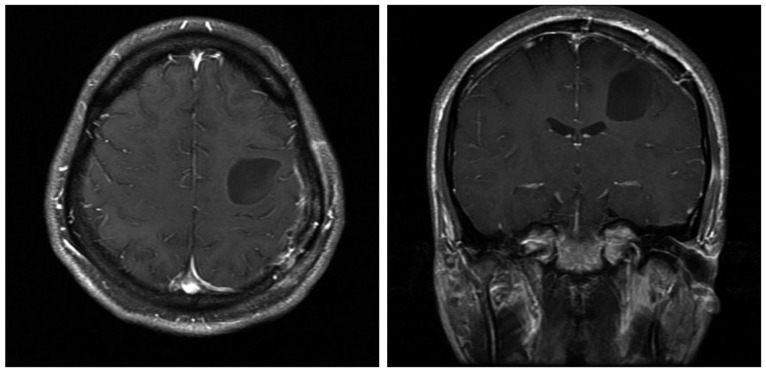
Fig. 4 One month after the operation, postoperative T1 enhance images shows no residual enhancing mass. The size of the peritumoral cyst is decreased, but remains after the operation.

Fig. 5 A : The tumor shows a fascicular arrangement of spindle neoplastic cells with perivascular hyalinization of the blood vessel wall (H&E, ×100). B : The tumor cell nuclei and cytoplasm are robustly positive for S100 protein (S100 protein immunostaining, ×100).
Reference
-
1. Barnard ZR, Agarwalla PK, Jeyaretna DS, Farrell CJ, Gerstner ER, Tian D, et al. Sporadic primary malignant intracerebral nerve sheath tumors : case report and literature review. J Neurooncol. 2011; 104:605–610. PMID: 21327709.
Article2. Bristol RE, Coons SW, Rekate HL, Spetzler RF. Invasive intracerebral schwannoma mimicking meningioma in a child. Childs Nerv Syst. 2006; 22:1483–1486. PMID: 17021734.
Article3. Bruni P, Esposito S, Greco R, Oddi G. Solitary intracerebral schwannoma in von Recklinghausen's disease. Surg Neurol. 1984; 22:360–364. PMID: 6433498.
Article4. Casadei GP, Komori T, Scheithauer BW, Miller GM, Parisi JE, Kelly PJ. Intracranial parenchymal schwannoma. A clinicopathological and neuroimaging study of nine cases. J Neurosurg. 1993; 79:217–222. PMID: 8331403.5. Cervoni L, Caruso R, Gagliardi FM. Intracerebral schwannoma. Case report. J Neurosurg Sci. 1998; 42:57–59. PMID: 9766275.6. Ghatak NR, Norwood CW, Davis CH. Intracerebral schwannoma. Surg Neurol. 1975; 3:45–47. PMID: 1111146.7. Gibson AA, Hendrick EB, Conen PE. Case reports. Intracerebral schwannoma. Report of a case. J Neurosurg. 1966; 24:552–557. PMID: 5935382.8. Guha D, Kiehl TR, Krings T, Valiante TA. Intracerebral schwannoma presenting as classic temporal lobe epilepsy. J Neurosurg. 2012; 117:136–140. PMID: 22559850.
Article9. Huang PP, Zagzag D, Benjamin V. Intracranial schwannoma presenting as a subfrontal tumor: case report. Neurosurgery. 1997; 40:194–197. PMID: 8971843.
Article10. Jung JM, Shin HJ, Chi JG, Park IS, Kim ES, Han JW. Malignant intraventricular schwannoma. Case report. J Neurosurg. 1995; 82:121–124. PMID: 7815115.11. Menkü A, Oktem IS, Kontas O, Akdemir H. Atypical intracerebral schwannoma mimicking glial tumor: case report. Turk Neurosurg. 2009; 19:82–85. PMID: 19263360.12. Oztanir N, Emmez H, Aytar MH, Dogan M, Kaymaz M, Baykaner MK. Malignant intracerebral giant nerve sheath tumor in a 14-month-old girl with neurofibromatosis type 1 : a case report. Childs Nerv Syst. 2009; 25:253–256. PMID: 18972118.
Article13. Sharma MC, Karak AK, Gaikwad SB, Mahapatra AK, Mehta VS, Sudha K. Intracranial intraparenchymal schwannomas : a series of eight cases. J Neurol Neurosurg Psychiatry. 1996; 60:200–203. PMID: 8708655.14. Takei H, Schmiege L, Buckleair L, Goodman JC, Powell SZ. Intracerebral schwannoma clinically and radiologically mimicking meningioma. Pathol Int. 2005; 55:514–519. PMID: 15998381.
Article15. Zagardo MT, Castellani RJ, Rees JH, Rothman MI, Zoarski GH. Radiologic and pathologic findings of intracerebral schwannoma. AJNR Am J Neuroradiol. 1998; 19:1290–1293. PMID: 9726470.
- Full Text Links
-
- Actions
-
Cited
- CITED
-
- Close
- Share
-
- Similar articles
-
- Supratentorial Intracerebral Neuroepithelial Cyst: Case Report
- Surgical Management of the Hypertensive Intracerebral Hemorrhage
- Remote Intracerebral Hematoma after Supratentorial Graniotomy
- Clinical Study on the Supratentoial Arteriovenous Malformation
- Efficacy of Percutaneous Needle Aspiration for Evacuation of Intracerebral Hemorrhage: A Volumetric and Clincal Outcone Study
