Infect Chemother.
2009 Oct;41(5):293-297. 10.3947/ic.2009.41.5.293.
Acute Pyelonephritis Complicated by Renal Vein Thrombosis and Pulmonary Embolism in Patient without any Thrombotic Risks
- Affiliations
-
- 1Department of Internal Medicine, Inha University School of Medicine, Incheon, Korea. retitia@daum.net
- 2Department of General Surgery, Inha University School of Medicine, Incheon, Korea.
- 3Department of Radiology, Inha University School of Medicine, Incheon, Korea.
- KMID: 1473656
- DOI: http://doi.org/10.3947/ic.2009.41.5.293
Abstract
- Renal vein thrombosis (RVT) is not an uncommon condition amongst patients with nephrotic syndrome or malignancy. Septic pulmonary embolism (SPE) is associated with risk factors such as intravenous drug use, pelvic thrombophlebitis, and suppurative processes in the head and neck. However, acute pyelonephritis is a rare cause of RVT and SPE. Case reports on RVT and SPE due to acute pyelonephritis are rare. In most of the earlier cases, patients had underlying conditions such as diabetes mellitus, renal carcinoma, calyceal stones, and hyperhomocysteinemia. We report a case of acute pyelonephritis complicated by RVT and SPE that occurred in a patient without any predisposing risk factors for thromboembolism. RVT and SPE were diagnosed using computed tomography and ventilation/perfusion scan. The patient recovered with antibiotics and anticoagulation therapy without any surgical interventions.
MeSH Terms
Figure
-

Figure 1 Chest x-ray shows small amount of pleural effusion and multiple nodular lesions in both lung fields (A). Abdominal CT shows low attenuated lesion in upper pole of right kidney (B lower arrow) and thrombosis in right renal vein (B upper arrow).
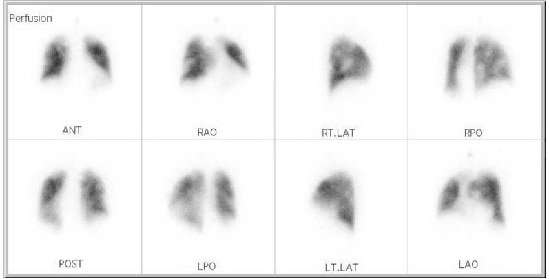
Figure 2 Ventilation-perfusion scan of the lung shows multiple nonsegmental perfusion defects.
Reference
-
1. Rossi SE, Goodman PC, Franquet T. Nonthrombotic pulmonary emboli. AJR Am J Roentgenol. 2000. 174:1499–1508.
Article2. Griffith GL, Maull KI, Sachatello CR. Septic pulmonary embolization. Surg Gynecol Obstet. 1977. 144:105–108.3. MacMillan JC, Milstein SH, Samson PC. Clinical spectrum of septic pulmonary embolism and infarction. J Thorac Cardiovasc Surg. 1978. 75:670–679.
Article4. O'Donnell AE, Selig J, Aravamuthan M, Richardson MS. Pulmonary complications associated with illicit drug use: an update. Chest. 1995. 108:460–463.5. Cook RJ, Ashton RW, Aughenbaugh GL, Ryu JH. Septic pulmonary embolism: presenting features and clinical course of 14 patients. Chest. 2005. 128:162–166.6. MacMillan JC, Milstein SH, Samson PC. Clinical spectrum of septic pulmonary embolism and infarction. J Thorac Cardiovasc Surg. 1978. 75:670–679.
Article7. Eijsten A, Leisinger HJ, Jucker A. Unilateral pyonephrosis with septic thrombosis of the renal vein and vena cava. Urol Int. 1986. 41:77–79.
Article8. Mamzer-Bruneel MF, Anglicheau D, Correas JM, Skhiri H, Jacobs F, Chrétien Y, Legendre C, Kreis H. Renal venous thrombosis: a forgotten complication of acute pyelonephritis. Presse Med. 1997. 26:1334–1336.9. Choi MJ, Kim TH, Lee JD, Cho YB, Lee HS, Jeon JS, Min HJ, Lee JG, Song CS. A case of acute pye lonephritis complicated with renal vein thrombosis. Korean J Med. 2000. 59:339–342.10. Kumar S, Singh SK, Mavuduru RS, Acharya NC, Agarwal MM, Jha VK, Mandal AK. Acute pyelonephritis with renal vein and inferior vena cava thrombosis in a case of hyperhomocysteinemia. Int Urol Nephrol. 2009. 41:185–188.
Article11. Lee SJ, Cha SI, Kim CH, Park JY, Jung TH, Jeon KN, Kim GW. Septic pulmonary embolism in Korea: Microbiology, clinicoradiologic features, and treatment outcome. J Infect. 2007. 54:230–234.
Article12. Günalp M, Gürler S, Polat O, Demirkan A. Septic pulmonary embolism associated with renal abscess: a case report. J Emerg Med. Forthcoming 2009.
Article13. Lee HY, Kim NH, Kim JH, Kim DH, Kim YS, Jee YK, Park JS. A case of septic pulmonary embolism due to pyelonephritis. Korean J Med. 2009. 76:105–109.14. Zenda T, Araki I, Hiraiwa Y, Miyayama S, Masunaga T, Takeda Y, Ueno T, Takeda R. Septic pulmonary emboli secondary to pyogenic liver abscess in a diabetic patient. Intern Med. 1995. 34:42–45.
Article15. Tsai AW, Cushman M, Rosamond WD, Heckbert SR, Polak JF, Folsom AR. Cardiovascular risk factors and venous thromboembolism incidence: the longitudinal investigation of thromboembolism etiology. Arch Intern Med. 2002. 162:1182–1189.
Article16. Ageno W, Becattini C, Brighton T, Selby R, Kamphuisen PW. Cardiovascular risk factors and venous thromboembolism: a meta-analysis. Circulation. 2008. 117:93–102.
Article17. Sakuma M, Sugimura K, Nakamura M, Takahashi T, Kitamukai O, Yazu T, Yamada N, Ota M, Kobayashi T, Nakano T, Shirato K. Unusual pulmonary embolism: septic pulmonary embolism and amniotic fluid embolism. Circ J. 2007. 71:772–775.
Article18. Lederman ER, Crum NF. Pyogenic liver abscess with a focus of Klebsiella pneumoniae as a primary pathogen: an emerging disease with unique clinical characteristics. Am J Gastroenterol. 2005. 100:322–331.
Article19. Golpe R, Marín B, Alonso M. Lemierre's syndrome (necrobacillosis). Postgrad Med J. 1999. 75:141–144.
- Full Text Links
-
- Actions
-
Cited
- CITED
-
- Close
- Share
-
- Similar articles
-
- A Case of Emphysematous Pyelonephritis Complicated with Septic Pulmonary Embolism
- A case of septic pulmonary embolism due to pyelonephritis
- A Case of Acute Pyelonephritis Complicated with Renal Vein Thrombosis
- A case of acute pyelonephritis complicated with renal vein thrombosis
- An Acute Pulmonary Embolism Accompanying Greater Saphenous Vein Thrombosis
