Diverse Characteristics of Spinal Nerve Sheath Tumor on Magnetic Resonance Images
- Affiliations
-
- 1Department of Orthopedics, Chonnam National University Hospital, Gwangju, Korea. hjkim2016@gmail.com
- KMID: 1473388
- DOI: http://doi.org/10.4184/jkss.2009.16.1.38
Abstract
-
STUDY DESIGN: A restrosepctive study
OBJECTIVES
We present the diverse imaging features of a spinal nerve sheath tumor for the preoperative diagnosis and treatment. SUMMARY OF LITERATURE REVIEW: The typical imaging findings of spinal nerve sheath tumors are reported in the literature. However, they can show diverse and unusual imaging features.
MATERIALS AND METHODS
The study group consisted of 30 patients who had undergone MR imaging for a preoperative evaluation of a spinal nerve sheath tumor from September 1989 to February 2008. All patients had undergone surgery for a spinal tumor that was confirmed by biopsy. The mean follow-up period was 13.1 months. The T1-, T2-weighted spine echo images and contrast material images were obtained in the sagittal plane. Axial images were obtained in any area of the spine where the sagittal images demonstrated abnormal findings. The signal intensity of the lesion, homogenicity, heterogenicity were evaluated in the T1-, T2-, and enhanced images
RESULTS
Twenty-four cases were neurilemmoma and 6 cases were neurofibromas. Different types of neurilemmomas included neurilemmomas with cystic changes (n=6), focal hemorrhage (n=5), extensive vertebral destruction (n=1), and giant neurilemmoma(n=1). The T1-weighted image showed low and intermediate signal intensity. The T2-weighted image showed high-signal intensity except for one neurilemmoma. The Gd-DTPA enhanced image showed homogenous, heterogeneous, and rim enhancement except for one case of a neurilemmoma with cystic changes.
CONCLUSIONS
Spinal nerve sheath tumors can show diverse and unusual imaging findings. An awareness of the uncommon presentations of these tumors is important for making a preoperative diagnosis and treatment. MRI is valuable in characterizing the soft tissue and bony anatomy in spinal neurilemmoma and neurofibroma.
Keyword
MeSH Terms
Figure
-

Fig. 1. Large or giant neurilemmoma. Sagittal T1 (A), T2 (B), and postcontrast (C) image show a large neurilemmoma extending more than 2 vertebral level (L5-S1). T1-weighted image (A) show low signal intensity and T2-weighted image (B) show high signal intensity.

Fig. 2. Neurilemmoma with extensive vertebral destruction. Sagittal T1 (A) and T2 (B) image show neurilemmoma involving vertebral body pedicle. Axial enhance (C) image and CT (D) show vertebral body with scalloping of its posterior border and neurilemmoma displacing the spinal cord.
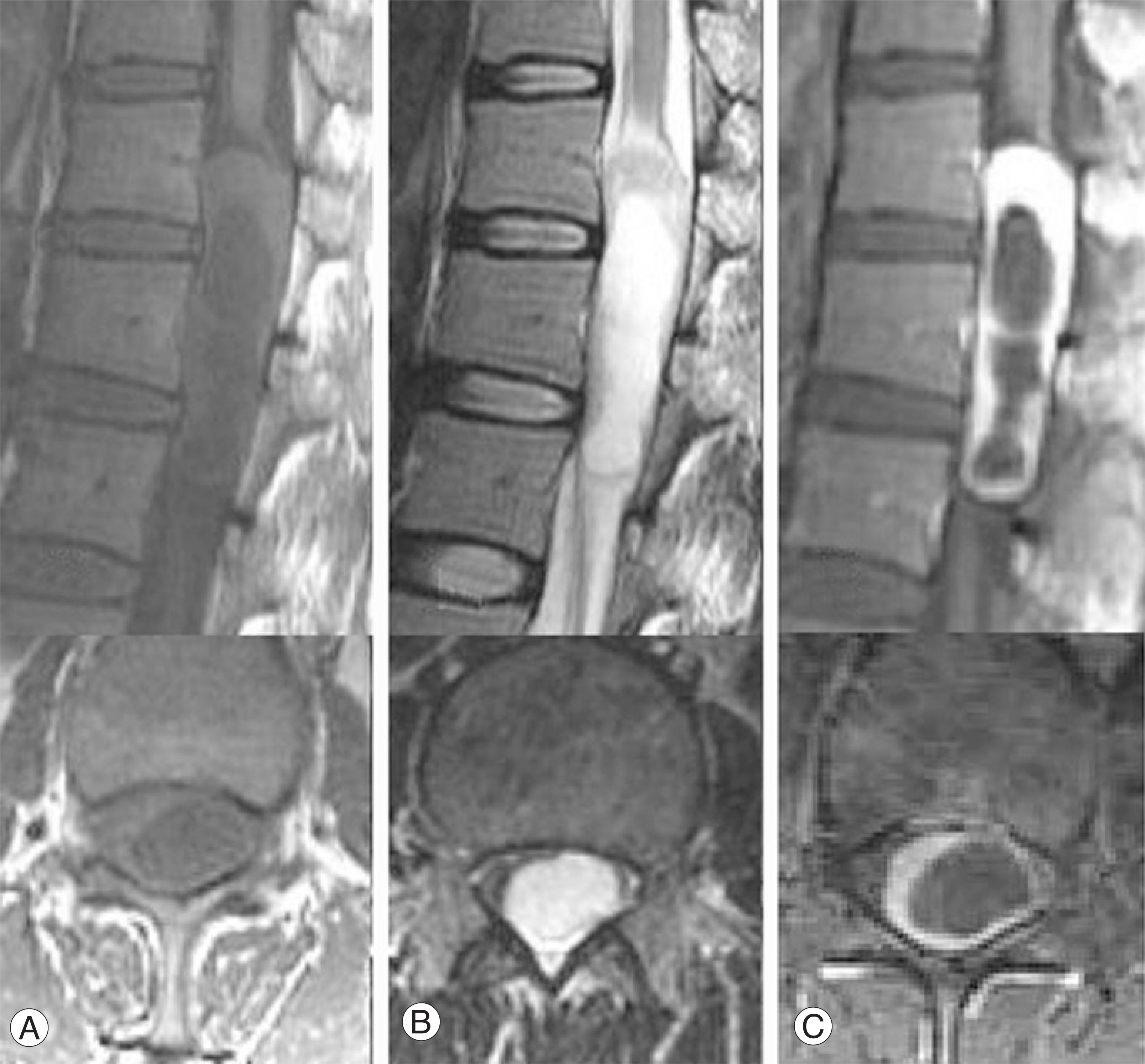
Fig. 3. Neurilemmoma with cystic changes. Sagittal and axial T1 (A),T2 (B), and postcontrast (C) image show a cystic intradural neurilemmoma at thoracic spine. The tumor shows peripherally enhancing margin with nodularity and different thickness of cyst wall.
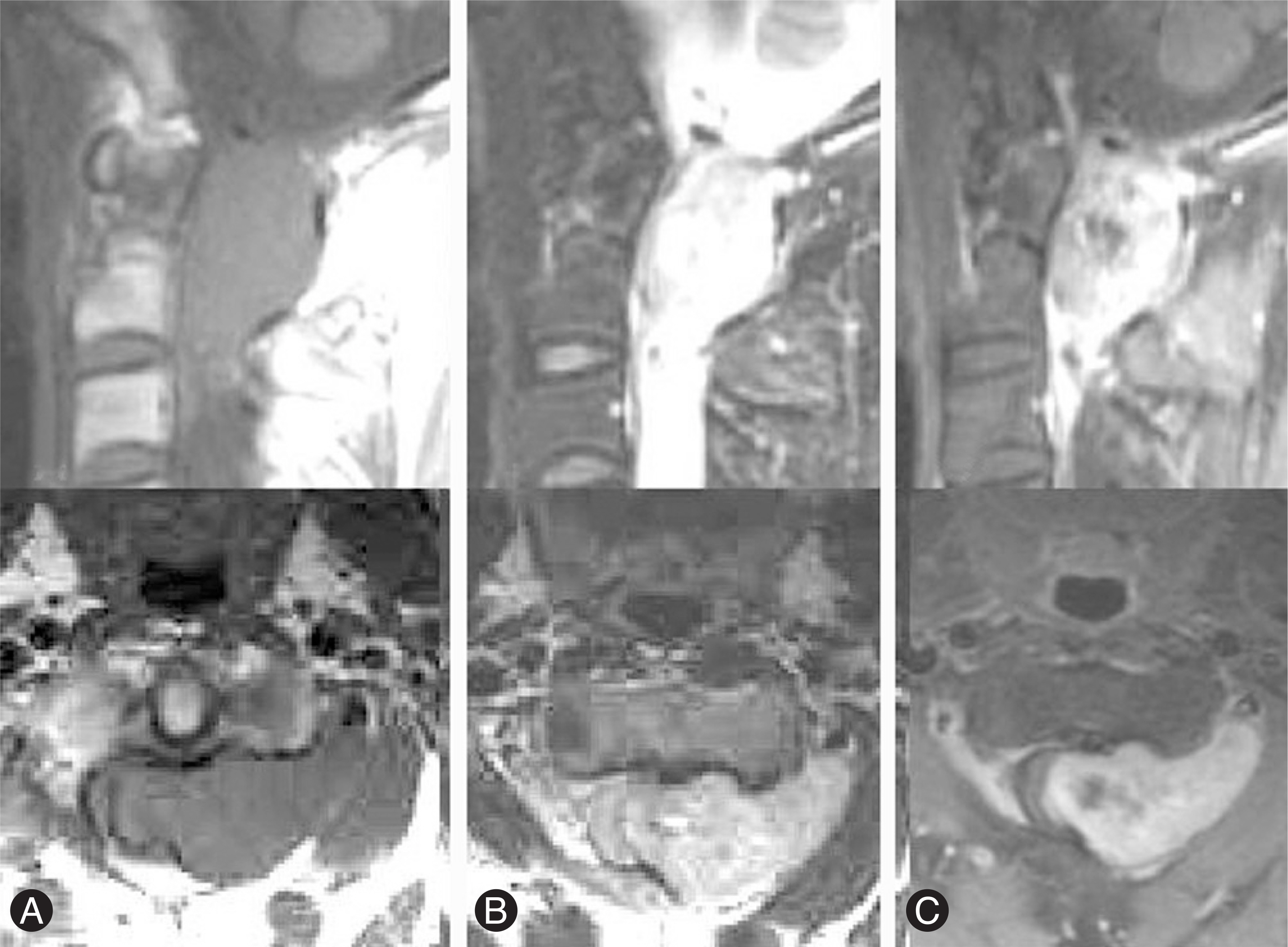
Fig. 4. Neurilemmoma with focal hemorrhage. Sagittal and axial T1 (A), T2 (B) image show a dumbbell shape neurilemmoma.with extraforaminal soft tissue extension. Postcontrast (C) image show centrally nonenhacing neurilemmoma. T1-weighted image (A) show intermediate signal intensity and T2-weighted image (B) show high signal intensity.
Cited by 1 articles
-
Synchronous Development of Spinal Cord Tumor: Meningioma and Schwannoma - A Case Report -
Hak-Jin Min, Jin-Soo Kim, Ju-Pil Seok
J Korean Soc Spine Surg. 2011;18(4):263-267. doi: 10.4184/jkss.2011.18.4.263.
Reference
-
1). Osborn AG. Intradural extramedullary tumors, cyst, and tumor like masses. Diagnostic neuroradiology, 2nd. St Louis, Mosby;895. 1994.2). Friedman DP., Tartaglino LM., Flanders AE. Intradural schwannomas of the spine: MR findings with emphasis on contrast-enhancement characteristics. AJR. 1992. 158:1347–1350.
Article3). Demachi H., Takashima T., Kadoya M, et al. MR imaging of spinal neurinomas with pathologic correlation. J Comput Assist Tomogr. 1990. 14:250–254.4). Bloomer CW., Ackerman A., Bhatia RG. Imaging for spinal tumors and new applications. Top Magn Reson Imaging. 2006. 17:69–87.5). Parmar H., Patkar D., Gadani S., Shah J. Cystic lumbar nerve sheath tumors: MRI features in 5 patients. Australas Radiol. 2001. 45:123–127.6). Parmar H., Pang BC., Lim CC., Chng SM., Tan KK. Spinal schwannoma presenting with acute subarachnoid hemorrhage: a diagnostic challenge. Am J Neuroradiol. 2004. 25:846–850.7). Buhl R., Barth H., Hugo HH., Mautner VF., Mehdorn HM. Intracranial and spinal melanotic schwannoma in the same patients. J Neurooncol. 2004. 68:249–254.8). Inaoka T., Takahashi K., Hanaoka H, et al. Paravertebral neurinoma associated with aggressive intervertebral extension. Skeletal Radiol. 2001. 30:286–289.9). Sridhar K., Ramamurthi R., Vasudevan MC., Rama-murthi B. Giant invasive spinal schwannomas: definition and surgical management. J Neurosurg. 2001. 94:210–215.
Article
- Full Text Links
-
- Actions
-
Cited
- CITED
-
- Close
- Share
-
- Similar articles
-
- Sequestered Disc Mimicking Benign Neurogenic Tumor: Report of 2 Cases
- Malignant Peripheral Nerve Sheath Tumor of Non-Neurofibromatosis Type I Metastasized to the Cerebrospinal Axis
- Optic nerve sheath meningioma mimicking optic perineuritis
- The Early Detection of Recurrence of Malignant Peripheral Nerve Sheath Tumor by Frequent Magnetic Resonance Imaging
- Optic Nerve Sheath Meningocele with Optic Disc Pit
