Sequential Sacral Insufficiency Fracture After Unilateral Pubic Fractures: A Case Report
- Affiliations
-
- 1Department of Orthopaedic Surgery, Center for Joint Disease, Chonnam National University Hwasun Hospital, Jeonnam, Korea. tryoon@chonnam.ac.kr
- KMID: 1464235
- DOI: http://doi.org/10.11005/kjbm.2012.19.1.47
Abstract
- Osteoporotic fractures of the pelvic ring are not uncommon and among them sacral insufficiency fractures (SIFs) are often overlooked in patients with buttock or low back pain following no or minimal trauma, which results in diagnostic delays. SIFs are often bilateral and are commonly associated with other fractures - most frequently with a pubic ramus fracture. However, it remains unclear which fracture (sacral or pubic ramus) occurs first, and the only report on the subject found that the fracture sequence is initiated by a sacral fracture. The authors describe a case of sequential bilateral SIFs in a 74-year old woman following superior and inferior pubic rami fractures on one side. In conclusion we suggested that SIFs can occur after pelvic ring injury in any side because biomechanical disruption of the pelvic ring can induce the sacral fracture in patients with severe osteoporosis.
Figure
-
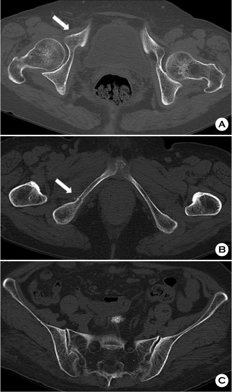
Fig. 1 Axial computed tomography image of the pelvis taken after a slip down showing a right superior (A) and an inferior (B) rami fracture (white arrow) but no fracture line on the sacrum (C).
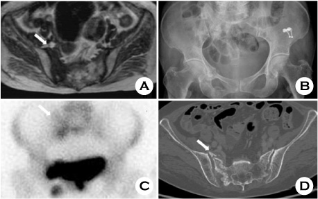
Fig. 2 (A) T1 weighted magnetic resonance imaging taken at the local hospital showing a low signal intensity lesion on the right sacral alar. (B) Anteroposterior radiograph of the pelvis showing right superior and inferior ramus callus formations and increased radiopaque on right sacral alar (white arrow). (C) Tc-99m bone scan showing increased uptakes at the right superior and inferior rami and right sacrum (white arrow). (D) Axial computed tomography image of the pelvis showing a right sacral fracture (white arrow). No fracture line was evident on the left sacrum.

Fig. 3 (A) Axial computed tomography (CT) image of the pelvis 2 months after the right sacral insufficiency fracture showing a new fracture line (white arrow). (B) Axial CT image of the pelvis 10 months after right sacral insufficiency fracture showing complete bone union of both sacral fractures.
Reference
-
1. Tsiridis E, Upadhyay N, Giannoudis PV. Sacral insufficiency fractures: current concepts of management. Osteoporos Int. 2006. 17:1716–1725.
Article2. Gotis-Graham I, McGuigan L, Diamond T, et al. Sacral insufficiency fractures in the elderly. J Bone Joint Surg Br. 1994. 76:882–886.
Article3. Grangier C, Garcia J, Howarth NR, May M, Rossier P. Role of MRI in the diagnosis of insufficiency fractures of the sacrum and acetabular roof. Skeletal Radiol. 1997. 26:517–524.
Article4. Weber M, Hasler P, Gerber H. Insufficiency fractures of the sacrum. Twenty cases and review of the literature. Spine (Phila Pa 1976). 1993. 18:2507–2512.5. De Smet AA, Neff JR. Pubic and sacral insufficiency fractures: clinical course and radiologic findings. AJR Am J Roentgenol. 1985. 145:601–606.
Article6. Lourie H. Spontaneous osteoporotic fracture of the sacrum. An unrecognized syndrome of the elderly. JAMA. 1982. 248:715–717.
Article7. Schindler OS, Watura R, Cobby M. Sacral insufficiency fractures. J Orthop Surg (Hong Kong). 2007. 15:339–346.
Article8. Linstrom NJ, Heiserman JE, Kortman KE, et al. Anatomical and biomechanical analyses of the unique and consistent locations of sacral insufficiency fractures. Spine (Phila Pa 1976). 2009. 34:309–315.
Article9. Pommersheim W, Huang-Hellinger F, Baker M, Morris P. Sacroplasty: a treatment for sacral insufficiency fractures. AJNR Am J Neuroradiol. 2003. 24:1003–1007.10. Tsiridis E, Upadhyay N, Gamie Z, Giannoudis PV. Percutaneous screw fixation for sacral insufficiency fractures: a review of three cases. J Bone Joint Surg Br. 2007. 89:1650–1653.
- Full Text Links
-
- Actions
-
Cited
- CITED
-
- Close
- Share
-
- Similar articles
-
- A Case of S1 Radiculopathy in Sacral Insufficiency Fracture without Fracture Line
- Usefulness of Kyphoplasty in Sacral Insufficiency Fracture: A Case Report
- MR Findings of Sacral Insufficiency Fractures in Osteoporotic Patients: Two Cases Report
- Sacral Insufficiency Fracture, Usually Overlooked Cause of Lumbosacral Pain
- Clinical observation of 62 of the pelvic bone fractures
