Arthroscopy of the Hip Joint: Diagnosis and Treatment
- Affiliations
-
- 1Department of Orthopaedic Surgery, School of Medicine, Chungnam National University, Daejeon, Korea. dshwang@cnu.ac.kr
- 2Department of Orthopaedic Surgery, College of Medicine, Konyang University, Daejeon, Korea.
- KMID: 1461074
- DOI: http://doi.org/10.5371/jkhs.2010.22.1.27
Abstract
- Hip arthroscopy is technically demanding and requires special distraction tools and operating equipment. The indications for hip arthroscopy are expanding as the understanding of hip disease increases. Improved instrumentation and technical skills have also facilitated the ability of physicians to treat some hip disorders arthroscopically. Various arthroscopic techniques allow the treatment of labral and acetabular rim pathology as well as pathology of the peripheral compartment. Improved techniques and longer-term outcomes studies should further define the optimal role of hip arthroscopy. Consequently, hip arthroscopy has been used to treat patients who should have had a more complicated open procedure or should have gone untreated. Moreover, hip arthroscopy, as with any procedure, is not without risks. Fortunately, complications are few, occurring in <5% of patients with hip pain.
Keyword
MeSH Terms
Figure
-
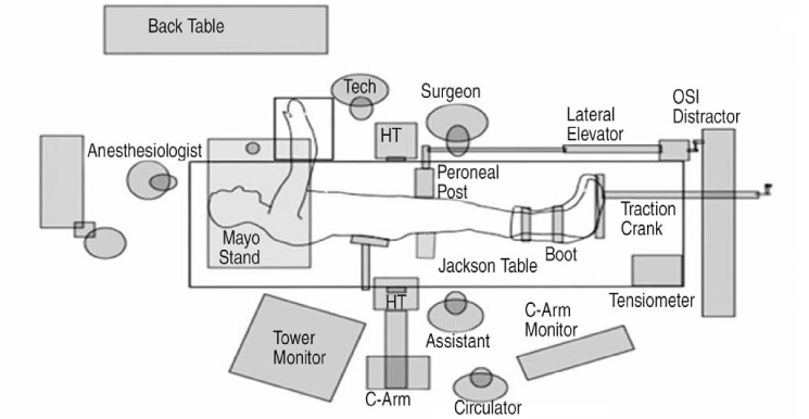
Fig. 1 Schematics of the operating room layout in lateral position.
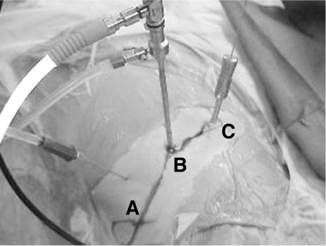
Fig. 2 Commonly used portals in lateral approach. (A) Proximal trochanteric, (B) Anterior paratrochanteric, (C) Posterior paratrochanteric
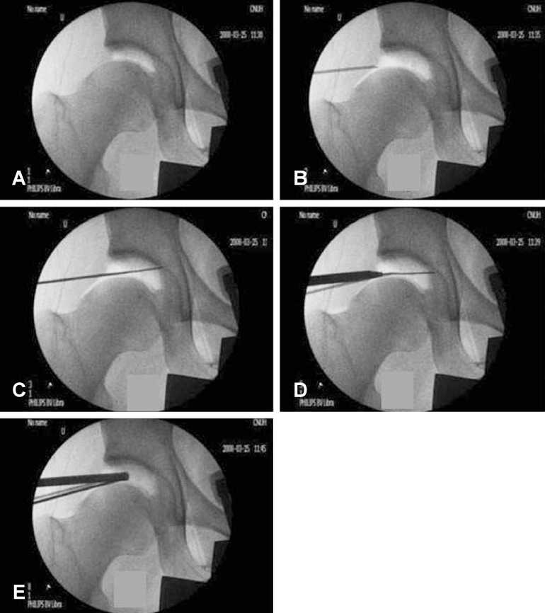
Fig. 3 Serial fluoroscopic images of classic portal insertion method in central compartment. (A) Vacuum seal shadow due to the negative intracapsular pressure is created by distraction of the joint. (B) The needle is inserted and the stylus is removed, then break the seal (C) as in air arthrogram, confirm the lateral labral silhouette. (D) The cannula/obturator assembly is being passed over the guide wire that had been placed through the spinal needle. (E) Then anterior portal is made.
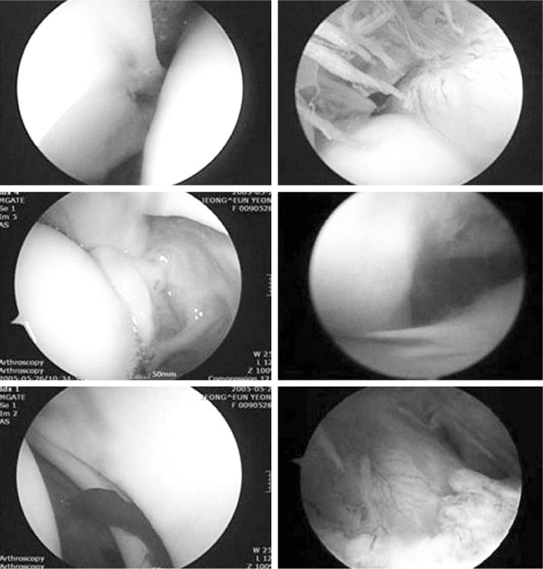
Fig. 4 Two compartments (central and peripheral) and normal arthroscopic findings. Acetabular labrum is the boundary to divide the two compartments.
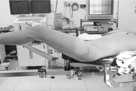
Fig. 5 Non-traction technique in hip arthroscopy.
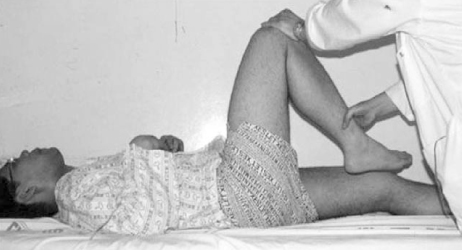
Fig. 6 Anterior impingement test. Affected hip is flexed more than 90 degree and adducted, internal rotated maneuver can cause anterior groin pain.
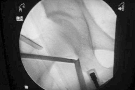
Fig. 7 Fluoroscopic image of medial approach.
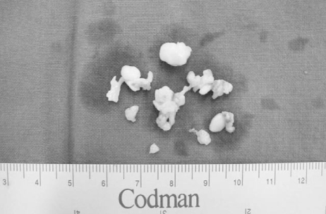
Fig. 8 Osteochondral loose bodies extracted by arthroscopic technique.
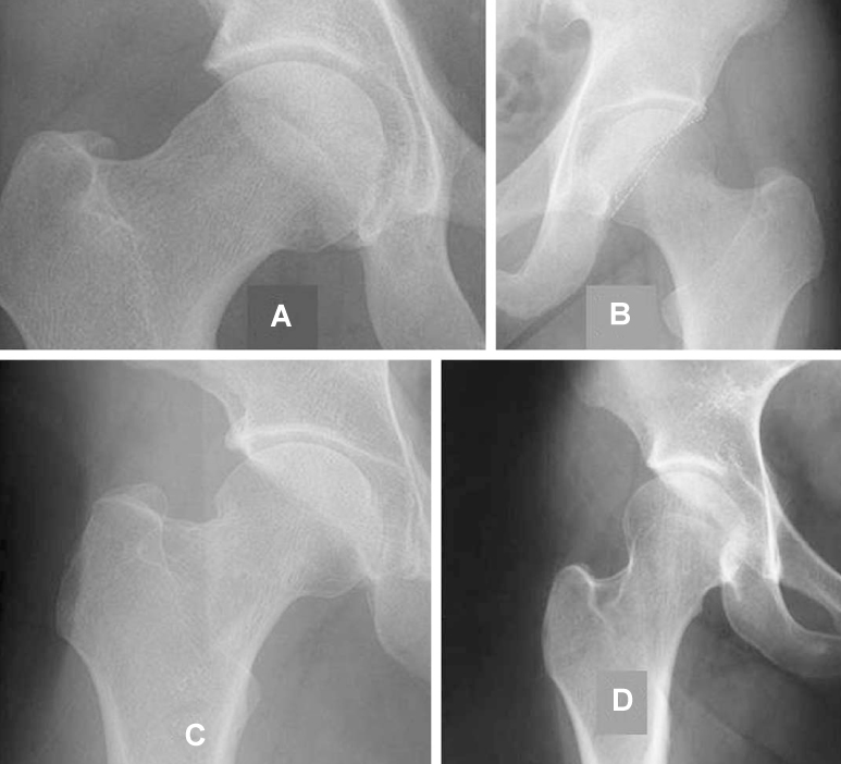
Fig. 9 Osseous abnormalities resulted in FAI. (A) Pistol grip deformity, (B) Retroverted acetabuli, (C) Coxa vara, (D) Coxa valga.
Reference
-
1. Burman MS. Arthroscopy or the direct visualization of joints. An experimental cadaver study. J Bone Joint Surg Am. 1931. 13:669–695.2. Takagi K. The arthroscope:the second report. J Jpn Orthop Assoc. 1939. 14:441–466.3. Gross RH. Arthroscopy in hip disorders in children. Orthop Rev. 1977. 6:43–49.4. Johnson LL. Diagnostic and surgical arthroscopy: The knee and other Joints. 1981. 2nd ed. St. Louis: Mosby.5. Watanabe M, Takeda S, Ikeuchi H. Atlas of Arthroscopy. 1969. 2nd ed. Tokyo: Igaku-shoin.6. Eriksson E, Arvidsson I, Arvidsson H. Diagnostic and operative arthroscopy of the hip. Orthopedics. 1986. 9:169–176.
Article7. Glick JM, Sampson TG, Gordon BB, Behr JT, Schmidt E. Hip arthroscopy by the lateral approach. Arthroscopy. 1986. 3:4–12.
Article8. Sampson TG. Complications of hip arthroscopy. Clin Sports Med. 2001. 20:831–835.
Article9. Villar RN. Arthroscopic debridement of the hip. J Bone Joint Surg Br. 1991. 73:suppl. 170–171.10. Villar RN. Hip Arthrocopy. 1992. Oxford: Butterworth-Heinemann.11. Conn KS, Villar RN. Labrum lesions from the viewpoint of arthroscopic hip surgery. Orthopade. 1998. 27:699–703.12. Dameron TB. Bucket-handle tears of acetabular labrum accompanying posterior dislocation of the hip. J Bone Joint Surg Am. 1959. 41:131–134.
Article13. Petersilege CA, Haque MA, Petersilge WJ, Lewin JS, Lieberman JM, Buly R. Acetabular labral tears: evaluation with MR arthrography. Radiology. 1996. 200:231–235.
Article14. Santori N, Villar RN. Acetabular labral tears: result of arthroscopic partial limbectomy. Arthroscopy. 2000. 16:11–15.
Article15. Okada Y, Awaya G, Ikeda T, Tada H, Kamisato S, Futami T. Arthroscopic surgery for synovial chondromatosis of the hip. J Bone Joint Surg Br. 1989. 71:198–199.
Article16. Hwang DS, Kim YM, Kim KC, Ahn SH. Arthroscopic treatment of acetabular labral tears. J Korean Hip Soc. 2004. 16:31–40.17. Dorfmann H, Boyer T. Arthroscopy of the hip: 12 years of experience. Arthroscopy. 1999. 15:67–72.
Article18. Glick JM. McGinty JH, editor. Hip Arthroscopy. Operative Arthroscopy. 1991. New York: Raven Press;663.19. Byrd JW. Hip arthroscopy. The supine position. Clin Sports Med. 2001. 20:703–731.20. McCarthy JC, Busconi B. The role of hip arthroscopy in the diagnosis and treatment of hip disease. Orthopedics. 1995. 18:753–756.
Article21. Keene GS, Villar RN. Arthroscopic anatomy of the hip: an in vivo study. Arthroscopy. 1994. 10:392–399.
Article22. McCarthy JC. Hip arthroscopy: Applications and technique. J Am Acad Orthop Surg. 1995. 3:115–122.
Article23. Fergurson SJ, Bryant JT, Ganz R, Ito K. The influence of the acetabular labrum on hip cartilage consolidation: a pororelastic finite element model. J Biomech. 2000. 33:953–960.24. Brumback RJ, Ellison TS, Molligan H, Molligan DJ, Mahaffey S, Schmidhauser C. Pudendal nerve palsy complicating intramedullary nailing of the femur. J Bone Joint Surg Am. 1992. 74:1450–1455.
Article25. Harris WH, Bourne RB, Oh I. Intra-articular acetabular labrum: a possible etiological factor in certain cases of osteoarthritis of the hip. J Bone Joint Surg Am. 1979. 61:510–514.26. Barlett CS, DiFelice GS, Buly RL, Quinn TJ, Green DS, Helfet DL. Cardiac arrest as a result of intraabdominal extravasations of fluid during arthroscopic removal of a loose body from the hip joint of a patient with an acetabular fracture. J Orthop Trauma. 1998. 12:294–299.27. Byrd JW. Byrd JWT, editor. The supine position. Operative Hip Arthroscopy. 1998. New York: Thieme;123–138.28. Dienst M, Seil R, Gödde S, Georg T, Kohn D. Arthroscopy for diagnosis and therapy of early osteoarthritis of the hip. Orthopade. 1999. 28:812–818.
Article29. Dienst M, Kohn D. Hip Arthroscopy. Minimal invasive diagnosis and therapy of the diseased or injured hip joint. Unfallchirurg. 2001. 104:2–18.30. Alterberg AR. Acetabular labrum tears: a cause of hip pain and degenerative arthritis. South Med J. 1977. 70:174–175.31. Fitzgerald RH Jr. Acetabular labrum tears. Diagnosis and treatment. Clin Orthop Relat Res. 1995. 311:60–68.32. MacDonald SJ, Klause K, Ganz R. The acetabular rim syndrome. Sem Arthroplast. 1997. 8:82–87.33. Leunig M, Werlin S, Ungersböck A, Ito K, Ganz R. Evaluation of the acetabular labrum by MR arthrography. J Bone Joint Surg Br. 1997. 79:230–234.
Article34. Czerny C, Hofmann S, Neuhold A, Tschauner C, Engel A, Recht MP, Kramer J. Lesions of the acetabular labrum : accuracy of MR imaging and MR arthrography in detection and staging. Radiology. 1996. 200:225–230.
Article35. Ruch DS, Satterfield W. The use of arthroscopy to document accurate position of core decompression of the hip. Arthroscopy. 1998. 14:617–619.
Article36. O'leary JA, Berend K, Vail TP. The relationship between diagnosis and outcome in arthroscopy of the hip. Arthroscopy. 2001. 17:181–188.37. Margheritini F, Villar RN. The efficacy of arthroscopy in the treatment of hip osteoarthritis. Chir Organi Mov. 1999. 84:257–261.38. Danielsson L, Lindberg H. Prevalence of coxarthrosis in an urban population during four decades. Clin Orthop Relat Res. 1997. 342:106–110.
Article39. Byrd JW. Labral lesions: an elusive source of hip pain case reports and literature review. Arthroscopy. 1996. 12:603–612.
Article40. Stiris MG. Magnetic resonance arthrography of the hip joint in patients with suspected rupture of labrum acetabulare. Tidsskr Nor Laegeforen. 2001. 121:698–700.41. Leuing M, Casillas MM, Halmet M, et al. Slipped capital femoral epiphysis: early mechanical damage to the acetabular cartilage by a prominent femoral metaphysis. Acta Ortho Scand. 2000. 71:370–375.
Article42. Ganz R, Parvizi J, Beck M, Leunig M, Nötzli H, Sienbenrock KA. Femoroacetabular impingement: a cause for osteoarthritis of the hip. Clin Orthop Relat Res. 2003. 417:112–120.43. Farjo LA, Glick JM, Sampson TG. Hip arthroscopy for acetabular labral tears. Arthroscopy. 1999. 15:132–137.
Article
- Full Text Links
-
- Actions
-
Cited
- CITED
-
- Close
- Share
-
- Similar articles
-
- Comprehensive Review of Advancements in Hip Arthroscopy
- Clinical Experiences of the Hip Arthroscopy
- Surgical Treatment of Acetabular Posterior Wall Fracture with Hip Arthroscopy: A Case Report
- The Current Concepts of Hip Arthroscopy
- Arthroscopic Removal of the Intraarticular Fracture Fragment after Internal Fixation of Acetabular Fracture: A Case Report
