Utility of the computed tomography indices on cone beam computed tomography images in the diagnosis of osteoporosis in women
- Affiliations
-
- 1Department of Oral and Maxillofacial Radiology, School of Dentistry, and Institute of Oral Bio Science, Chonbuk National University, Jeonju, Korea. kkj1512@jbnu.ac.kr
- KMID: 1449954
- DOI: http://doi.org/10.5624/isd.2011.41.3.101
Abstract
- PURPOSE
This study evaluated the potential use of the computed tomography indices (CTI) on cone beam CT (CBCT) images for an assessment of the bone mineral density (BMD) in postmenopausal osteoporotic women.
MATERIALS AND METHODS
Twenty-one postmenopausal osteoporotic women and 21 postmenopausal healthy women were enrolled as the subjects. The BMD of the lumbar vertebrae and femur were calculated by dual energy X-ray absorptiometry (DXA) using a DXA scanner. The CBCT images were obtained from the unilateral mental foramen region using a PSR-9000N(TM) Dental CT system. The axial, sagittal, and coronal images were reconstructed from the block images using OnDemend3D(TM). The new term "CTI" on CBCT images was proposed. The relationship between the CT measurements and BMDs were assessed and the intra-observer agreement was determined.
RESULTS
There were significant differences between the normal and osteoporotic groups in the computed tomography mandibular index superior (CTI(S)), computed tomography mandibular index inferior (CTI(I)), and computed tomography cortical index (CTCI). On the other hand, there was no difference between the groups in the computed tomography mental index (CTMI: inferior cortical width).
CONCLUSION
CTI(S), CTI(I), and CTCI on the CBCT images can be used to assess the osteoporotic women.
MeSH Terms
Figure
-
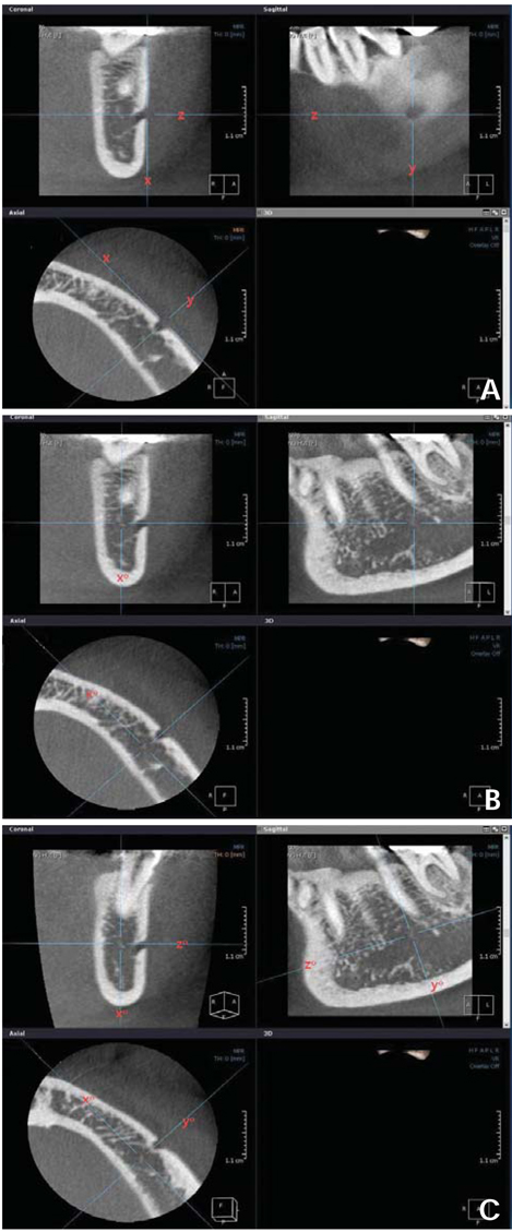
Fig. 1 A. On the axial image, an antero-posterior tangent line "x" is drawn, and line "y" and "z" are drawn perpendicular to line "x" through the center of the mental foramen, then these lines become perpendicular mutually. B. On the coronal image, line "x" is moved to line "x°", the bisecting position of the bucco-lingual dimension of the mandibular body. C. On the sagittal image, line "z" is moved to line "z°", parallel position with the mandibular inferior border of the mandible, then line "y" corresponds with line "y°", perpendicular line "z°" at the same time.

Fig. 2 A. Distance "S" from the superior margin of the mental foramen to the inferior border of the mandible is measured on coronal image. B. Distance "I" is measured from the inferior margin of the mental foramen to the inferior border of the mandible. C. Inferior cortical width (W) of the mandible is measured.
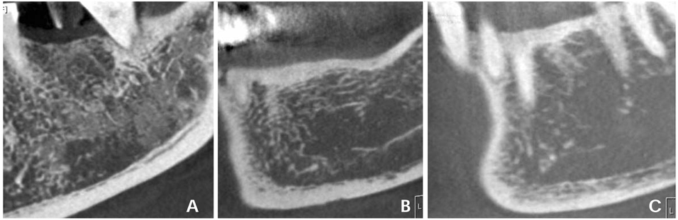
Fig. 3 The types of the inferior mandibular cortex are subjectively classified as follows. A. Type 1: the cortical endosteal margin appears even and regular. B. Type 2: the endosteal margin appears semilunar defects or 1 to 2 layers of cortical endosteal residues. C. Type 3: the cortical layer has numerous (>3) endosteal residues and is clearly porous.
Cited by 2 articles
-
Osteoporosis prediction from the mandible using cone-beam computed tomography
Imad Barngkgei, Iyad Al Haffar, Razan Khattab
Imaging Sci Dent. 2014;44(4):263-271. doi: 10.5624/isd.2014.44.4.263.IDIOS: An innovative index for evaluating dental imaging-based osteoporosis screening indices
Imad Barngkgei, Esam Halboub, Abeer Abdulkareem Almashraqi, Razan Khattab, Iyad Al Haffar
Imaging Sci Dent. 2016;46(3):185-202. doi: 10.5624/isd.2016.46.3.185.
Reference
-
1. Verheij JG, Geraets WG, van der Stelt PF, Horner K, Lindh C, Nicopoulou-Karayianni K, et al. Prediction of osteoporosis with dental radiographs and age. Dentomaxillofac Radiol. 2009. 38:431–437.
Article2. White SC. Oral radiographic predictors of osteoporosis. Dentomaxillofac Radiol. 2002. 31:84–92.
Article3. Nackaerts O, Jacobs R, Devlin H, Pavitt S, Bleyen E, Yan B, et al. Osteoporosis detection using intraoral densitometry. Dentomaxillofac Radiol. 2008. 37:282–287.
Article4. Kim JY, Nah KS, Jung YH. Comparison of panorama radiomorphometric indices of the mandible in normal and osteoporotic women. Korean J Oral Maxillofac Radiol. 2004. 34:69–74.5. Klemetti E, Kolmakov S, Heiskanen P, Vainio P, Lassila V. Panoramic mandibular index and bone mineral densities in postmenopausal women. Oral Surg Oral Med Oral Pathol. 1993. 75:774–779.
Article6. Hua Y, Nackaerts O, Duyck J, Maes F, Jacobs R. Bone quality assessment based on cone beam computed tomography imaging. Clin Oral Implants Res. 2009. 20:767–771.
Article7. Lindh C, Nilsson M, Klinge B, Petersson A. Quantitative computed tomography of trabecular bone in the mandible. Dentomaxillofac Radiol. 1996. 25:146–150.
Article8. Taguchi A, Tanimoto K, Suei Y, Ohama K, Wada T. Relationship between the mandibular and lumbar vertebral bone mineral density at different postmenopausal stages. Dentomaxillofac Radiol. 1996. 25:130–135.
Article9. Horner K, Karayianni K, Mitsea A, Berkas L, Mastoris M, Jacobs R, et al. The mandibular cortex on radiographs as a tool for osteoporosis risk assessment: the OSTEODENT project. J Clin Densitom. 2007. 10:138–146.
Article10. Karayianni K, Horner K, Mitsea A, Berkas L, Mastoris M, Jacobs R, et al. Accuracy in osteoporosis diagnosis of a combination of mandibular cortical width measurement on dental panoramic radiographs and a clinical risk index (OSIRIS): the OSTEODENT project. Bone. 2007. 40:223–229.
Article11. Horner K, Devlin H, Alsop CW, Hodgkinson IM, Adams JE. Mandibular bone mineral density as a predictor of skeletal osteoporosis. Br J Radiol. 1996. 69:1019–1025.
Article12. Taguchi A, Tsuda M, Ohtsuka M, Kodama I, Sanada M, Nakamoto T, et al. Use of dental panoramic radiographs in identifying younger postmenopausal women with osteoporosis. Osteoporos Int. 2006. 17:387–394.
Article13. Drozdzowska B, Pluskiewicz W, Tarnawska B. Panoramic-based mandibular indices in relation to mandibular bone mineral density and skeletal status assessed by dual energy X-ray absorptiometry and quantitative ultrasound. Dentomaxillofac Radiol. 2002. 31:361–367.
Article14. Horner K, Devlin H. The relationship between two indices of mandibular bone quality and bone mineral density measured by dual energy X-ray absorptiometry. Dentomaxillofac Radiol. 1998. 27:17–21.15. Taguchi A, Suei Y, Ohtsuka M, Otani K, Tanimoto K, Ohtaki M. Usefulness of panoramic radiography in the diagnosis of postmenopausal osteoporosis in women. Width and morphology of inferior cortex of the mandible. Dentomaxillofac Radiol. 1996. 25:263–267.
Article16. Ledgerton D, Horner K, Devlin H, Worthington H. Panoramic mandibular index as a radiomorphometric tool: an assessment of precision. Dentomaxillofac Radiol. 1997. 26:95–100.
Article17. Horner K, Allen P, Graham J, Jacobs R, Boonen S, Pavitt S, et al. The relationship between the OSTEODENT index and hip fracture risk assessment using FRAX. Oral Surg Oral Med Oral Pathol Oral Radiol Endod. 2010. 110:243–249.
Article18. Taguchi A, Suei Y, Sanada M, Ohtsuka M, Nakamoto T, Sumida H, et al. Validation of dental panoramic radiography measures for identifying postmenopausal women with spinal osteoporosis. AJR Am J Roentgenol. 2004. 183:1755–1760.
Article19. Adams JE. Osteoporosis and bone mineral densitometry. Curr Opin Radiol. 1992. 4:11–20.20. Benson BW, Prihoda TJ, Glass BJ. Variations in adult cortical bone mass as measured by a panoramic mandibular index. Oral Surg Oral Med Oral Pathol. 1991. 71:349–356.
Article21. Watson EL, Katz RV, Adelezzi R, Gift HC, Dunn SM. The measurement of mandibular cortical bone height in osteoporotic vs. non-osteoporotic postmenopausal women. Spec Care Dentist. 1995. 15:124–128.
Article22. Klemetti E, Kolmakov S, Kröger H. Pantomography in assessment of the osteoporosis risk group. Scand J Dent Res. 1994. 102:68–72.
Article23. Devlin H, Karayianni K, Mitsea A, Jacobs R, Lindh C, van der Stelt P, et al. Diagnosing osteoporosis by using dental panoramic radiographs: The OSTEODENT project. Oral Surg Oral Med Oral Pathol Oral Radiol Endod. 2007. 104:821–828.
Article24. Horner K, Devlin H. The relationship between mandibular bone mineral density and panoramic radiographic measurements. J Dent. 1998. 26:337–343.
Article
- Full Text Links
-
- Actions
-
Cited
- CITED
-
- Close
- Share
-
- Similar articles
-
- Three-dimensional imaging modalities in endodontics
- Management of root canal perforation by using cone-beam computed tomography
- Basic principle of cone beam computed tomography
- Detection of maxillary second molar with two palatal roots using cone beam computed tomography: a case report
- Foramen transversarium enlargement caused by vertebral artery tortuosity: Diagnosis with cone-beam computed tomography and magnetic resonance angiography
