Intraosseous ameloblastoma masquerading as exophytic growth: a case report
- Affiliations
-
- 1Department of Oral Medicine and Radiology, Dayananda Sagar College of Dental Sciences, Bangalore, India. sanjay_cj@yahoo.co.in
- 2Department of Oral Medicine and Radiology, Dr Shyamala Reddy Dental College and Hospital, Bangalore, India.
- KMID: 1449941
- DOI: http://doi.org/10.5624/isd.2011.41.2.89
Abstract
- Intraosseous ameloblastoma is the most common and simple type of ameloblastoma prevalent among odontogenic tumors. Clinico-radiographically intraosseous ameloblastoma presents as slow, painless swelling or expansion of the jaws and described as multilocular expansile radiolucency that occurs most frequently in mandibular molar/ramus area. This article describes a case of follicular ameloblastoma involving 45 year old male which is different from the usual presentation, which includes-exophytic growth, different location and without expansion of the cortex.
Keyword
MeSH Terms
Figure
-
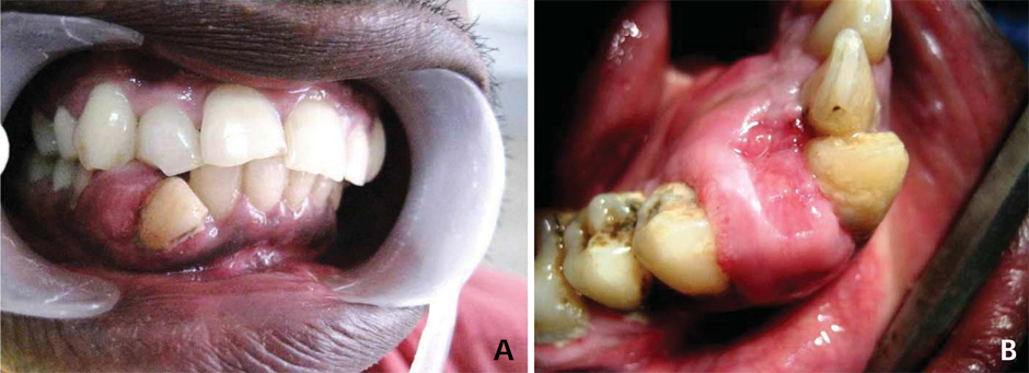
Fig. 1 Clinical photographs show an exophytic growth at the inter-dental area in right mandible between canine and first premolar extending labially (A) from the attached gingiva of the teeth to the lingual sulcus of incisors, canine, and first premolar teeth (B).
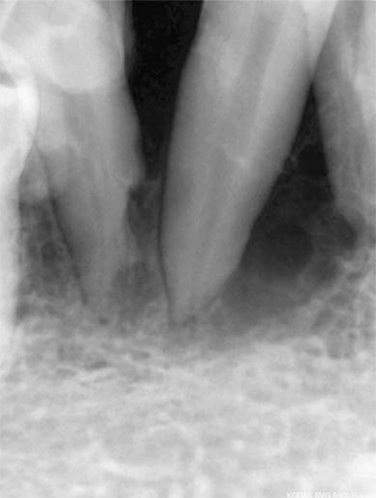
Fig. 2 Intraoral periapical radiograph shows a presence of characteristic soap bubble appearance in the region of the right mandibular canine and first premolar, and knife edge root resorption pattern in relation to the first premolar.
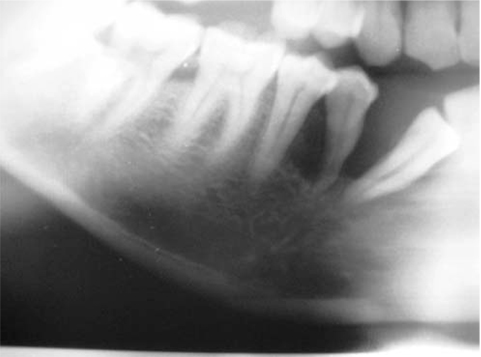
Fig. 3 Panoramic radiograph shows a radiolucent area measuring 4×4 cm in diameter extending from the distal aspect of the right lateral incisor to the mesial aspect of the second premolar.
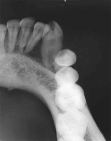
Fig. 4 Occlusal radiograph shows no cortical expansion.

Fig. 5 Gross specimen after surgical removal.
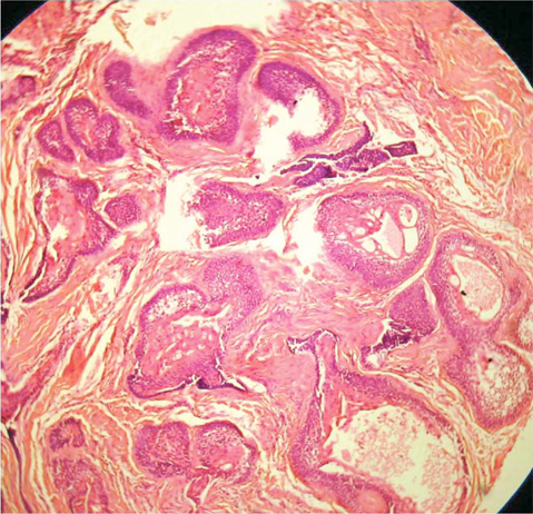
Fig. 6 Histopathological examination demonstrates connective tissue with numerous follicles lined by single layer of tall columnar ameloblast-like cells (H&E stain, 40×).
Reference
-
1. Langlais RP, Langland OE, Nortje CJ. Multilocular radiolucencies. Diagnostic imaging of jaws. 1995. 1st ed. Williams & Wilkins Co;327–384.2. Rajendran R. Rajendran R, Sivapathasundharam B, editors. Cyst and tumors of odntogenic origin. Shafer's text book of oral pathology. 2009. 6th ed. Noida: Elsevier;254–308.3. Philipsen HP, Reichart PA. Unicystic ameloblastoma. A review of 193 cases from the literature. Oral Oncol. 1998. 34:317–325.
Article4. Kim SG, Jang HS. Ameloblastoma: a clinical, radiographic, and histopathologic analysis of 71 cases. Oral Surg Oral Med Oral Pathol Oral Radiol Endod. 2001. 91:649–653.
Article5. Philipsen HP, Reichart PA, Nikai H, Takata T, Kudo Y. Peripheral ameloblastoma: biological profile based on 160 cases from the literature. Oral Oncol. 2001. 37:17–27.
Article6. Kuru H. Ueber das Adamantinom. Zentralbl Allg Pathol. 1911. 22:291–295.7. Tongdee C, Ganggavakin S. Peripheral ameloblastoma (report of a case and review literature). J Dent Assoc Thai. 1978. 28:31–38.8. Stevenson AR, Austin BW. A case of ameloblastoma presenting as an exophytic gingival lesion. J Periodontol. 1990. 61:378–381.
Article9. Ueno S, Nakamura S, Mushimoto K, Shirasu R. A clinicopathologic study of ameloblastoma. J Oral Maxillofac Surg. 1986. 44:361–365.
Article10. Waldron CA, el-Mofty SK. A histopathologic study of 116 ameloblastomas with special reference to the desmoplastic variant. Oral Surg Oral Med Oral Pathol. 1987. 63:441–451.
Article11. Eversole LR, Leider AS, Strub D. Radiographic characteristics of cystogenic ameloblastoma. Oral Surg Oral Med Oral Pathol. 1984. 57:572–577.
Article12. Martelli-Júnior H, Souza LN, Santos LA, Melo-Filho MR, De Paula AM. Peripheral ameloblastoma: a case report. Oral Surg Oral Med Oral Pathol Oral Radiol Endod. 2005. 99:E31–E33.
Article13. de Villiers-Slabbert H, Altini M. Peripheral odontogenic fibroma: a clinicopathologic study. Oral Surg Oral Med Oral Pathol. 1991. 72:86–90.14. Ide F, Kusama K, Tanaka A, Sakashita H. Peripheral ameloblastoma is not a hamartoma but rather more of a neoplasm. Oral Oncol. 2002. 38:318–320.
Article15. Yamamoto T, Ueta E, Yoneda K, Osaki T. Peripheral ameloblastoma: case report with immunohistochemical investigation. J Oral Maxillofac Surg. 1990. 48:197–200.
Article
