Management of white spots: resin infiltration technique and microabrasion
- Affiliations
-
- 1Department of Conservative Dentistry, Pusan National University School of Dentistry, Yangsan, Korea. jeongkil@pusan.ac.kr
- KMID: 1446085
- DOI: http://doi.org/10.5395/JKACD.2011.36.1.66
Abstract
- This case report compared the effectiveness of resin infiltration technique (Icon, DMG) with microabrasion (Opalustre, Ultradent Products, Inc.) in management of white spot lesions. It demonstrates that although neither microabrasion nor resin infiltration technique can remove white spot lesions completely, resin infiltration technique seems to be more effective than microabrasion. Therefore resin infiltration technique can be chosen preferentially for management of white spot lesions and caution should be taken for case selection.
Figure
-
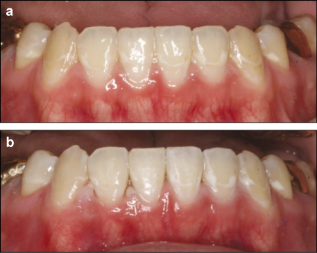
Figure 1
Intraoral photographs taken prior to treatment (a), after completion of resin infiltration technique (right side) and microabrasion (left side) (b). 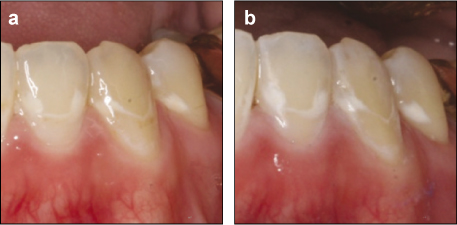
Figure 2
Photographs of left mandibular lateral incisor, canine, second premolar taken prior to treatment (a), after completion of microabrasion (b). 
Figure 3
Photographs of right mandibular lateral incisor, canine, second premolar taken prior to treatment (a), after completion of resin infiltration technique (b). 
Figure 4
Intraoral photographs taken prior to treatment (a) and after completion of resin infiltration technique (left maxillary central incisor), microabrasion (right maxillary central incisor) and resin composite restoration (right maxillary lateral incisor) (b). 
Figure 5
Photographs of maxillary central incisors taken prior to treatment (a) and completion of resin infiltration technique and microabrasion (b).
Cited by 3 articles
-
Non-destructive management of white spot lesions by using tooth jewelry
Hee-Jin Kim, Lorena Karanxha, Su-Jung Park
Restor Dent Endod. 2012;37(4):236-239. doi: 10.5395/rde.2012.37.4.236.Color and hardness changes in artificial white spot lesions after resin infiltration
Ji-Hoon Kim, Ho-Hyun Son, Juhea Chang
Restor Dent Endod. 2012;37(2):90-95. doi: 10.5395/rde.2012.37.2.90.Application of quantitative light-induced fluorescence to determine the depth of demineralization of dental fluorosis in enamel microabrasion: a case report
Tae-Young Park, Han-Sol Choi, Hee-Won Ku, Hyun-Su Kim, Yoo-Jin Lee, Jeong-Bum Min
Restor Dent Endod. 2016;41(3):225-230. doi: 10.5395/rde.2016.41.3.225.
Reference
-
1. Chang HS, Walsh LJ, Freer TJ. Enamel demineralization during orthodontic treatment. Aetiology and prevention. Aust Dent J. 1997. 42(5):322–327.
Article2. Kang HS, Lee CY. The influence of the degree of saturation of acidulated buffer solutions in the root dentin demineralization. J Korean Acad Conserv Dent. 2004. 29(5):454–461.
Article3. Sudjalim TR, Woods MG, Manton DJ. Prevention of white spot lesions in orthodontic practice: a contemporary review. Aust Dent J. 2006. 51(4):284–289.
Article4. Gorelick L, Geiger AM, Gwinnett AJ. Incidence of white spot formation after bonding and banding. Am J Orthod. 1982. 81(2):93–98.
Article5. Ogaard B, Rolla G, Arends J, ten Cate JM. Orthodontic appliances and enamel demineralization. Part 2. Prevention and treatment of lesions. Am J Orthod Dentofacial Orthop. 1988. 94(2):123–128.
Article6. Cha SW, Yoon TC, Park SH, Lee CY, Kum KY. Quantitative analysis of mineral change in the initial carious lesion using confocal laser scanning microscopy. J Korean Acad Conserv Dent. 2001. 26(1):1–8.7. Kwun JW, Kim KY, Lee SJ, Jung IY, Lee CY. The effect of the supersaturated solutions containing high concentrations of fluoride on seeded crystal growth. J Korean Acad Conserv Dent. 1999. 24(2):330–336.8. Willmot DR. White lesions after orthodontic treatment: does low fluoride make a difference? J Orthod. 2004. 31(3):235–242.
Article9. Al-Khateeb S, Extekate RA, de Josselin de Jong E, Angmar-Mansson B, ten Cate JM. Light-induced fluorescence studies on dehydration of incipient enamel lesions. Caries Res. 2002. 36(1):25–30.
Article10. Ardu S, Castiono NV, Benbachir N, Krejci I. Minimally invasive treatment of white spot enamel lesions. Quintessence Int. 2007. 38(8):633–636.11. Murphy TC, Willmote DR, Rodd HD. Management of postorthodontic demineralized white lesions with microabrasion: A quantitative assessment. Am J Orthod Dentofacial Orthop. 2007. 131(1):27–33.
Article12. Donly KJ, O'neill M, Croll TP. Enamel microabrasion: a microscopic evaluation of the "abrosion effect". Quintessence Int. 1992. 23(3):175–179.13. Tong LS, Pang MK, Mok NY, King NM, Wei SH. The effects of etching, micro-abrasion, and bleaching on surface enamel. J Dent Res. 1993. 72(1):67–71.
Article14. Paris S, Meyer-Lueckel H, Kielbassa AM. Resin infiltration of natural caries lesions. J Dent Res. 2007. 86(7):662–666.
Article15. Robinson C, Hallsworth AS, Weatherell JA, Kunzel W. Arrest and control of carious lesions: a study based on preliminary experiments with resorcinol-formaldehyde resin. J Dent Res. 1976. 55(5):812–818.
Article16. Kielbassa AM, Gernhardt CR. Closing the gap between oral hygiene and minimally invasive dentistry: A review on the resin infiltration technique of incipient (proximal) enamel lesions. Quintessence Int. 2009. 40(8):663–681.17. Kidd EA, Fejerskov O. What constitutes dental caries? Histopathology of carious enamel and dentin related to the action of cariogenic biofilms. J Dent Res. 2004. 83(Spec No C):C35–C38.
Article18. Paris S, Meyer-Lueckel H. Masking of labial enamel white spot lesions by resin infiltration - A clinical report. Quintessence Int. 2009. 40(9):713–718.19. Davila JM, Buonocore MG, Greeley CB, Provenza DV. Adhesive penetration in human artificial and natural white spots. J Dent Res. 1975. 54(5):999–1008.
Article
- Full Text Links
-
- Actions
-
Cited
- CITED
-
- Close
- Share
-
- Similar articles
-
- Non-destructive management of white spot lesions by using tooth jewelry
- Color and hardness changes in artificial white spot lesions after resin infiltration
- A global overview of enamel microabrasion for white spot lesions: a bibliometric review
- Application of quantitative light-induced fluorescence to determine the depth of demineralization of dental fluorosis in enamel microabrasion: a case report
- White spots on the mucosal surface of the duodenum in dogs with lymphocytic plasmacytic enteritis
