J Vet Sci.
2011 Jun;12(2):165-169. 10.4142/jvs.2011.12.2.165.
White spots on the mucosal surface of the duodenum in dogs with lymphocytic plasmacytic enteritis
- Affiliations
-
- 1Department of Animal Medicine and Surgery, College of Veterinary Medicine, Complutense University of Madrid, Avenida Puerta de Hierro s/n, 28040 Madrid, Spain. mercgarc@vet.ucm.es
- KMID: 1106559
- DOI: http://doi.org/10.4142/jvs.2011.12.2.165
Abstract
- Distended lacteals, described as expanded white villi in duodenum, are strongly indicative of primary intestinal lymphangiectasia. In the present study, we evaluated the significance of white spots present in the duodenal mucosa of dogs with lymphocytic plasmacytic enteritis (LPE). Fifty dogs with LPE were included in this study, and white spots were detected in the duodenal mucosa in 22 dogs during endoscopy. Hypoproteinemia was more frequent in dogs with white spots than in dogs without spots (p = 0.02). Serum protein and albumin concentration were significantly lower in LPE dogs with white spots (p = 0.038) compared to LPE dogs without white spots (p = 0.039). There was a significant correlation between white spots density and lymphatic dilatation histological scores (p = 0.023; rho = 0.481). These results suggest that the presence of white spots in the duodenal mucosa of dogs is not a finding exclusive for intestinal lymphangiectasia. Low serum protein and albumin concentrations together with lymphatic dilatation seem to be related to the presence of white spots in the duodenal mucosa of LPE dogs.
MeSH Terms
Figure
-
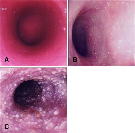
Fig. 1 Endoscopic appearance of duodenum showing white spots on the mucosal surface. (A) mild, (B) moderate, and (C) severe densities and distributions.
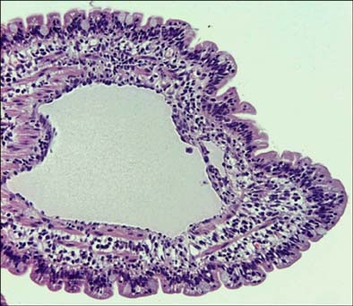
Fig. 2 Histopathological appearance of duodenum showing central lacteal ballooning that represented up to 75% of width of the villous lamina propria on longitudinal section. H&E stain, ×100.
Reference
-
1. Allenspach K, Wieland B, Gröne A, Gaschen F. Chronic enteropathies in dogs: Evaluation of risk factors for negative outcome. J Vet Intern Med. 2007. 21:700–708.
Article2. Burgener IA, König A, Allenspach K, Sauter SN, Boisclair J, Doherr MG, Jungi TW. Upregulation of toll-like receptors in chronic enteropathies in dogs. J Vet Intern Med. 2008. 22:553–560.
Article3. Craven M, Simpson JW, Ridyard AE, Chandler ML. Canine inflammatory bowel disease: retrospective analysis of diagnosis and outcome in 80 cases (1995-2002). J Small Anim Pract. 2004. 45:336–342.
Article4. Dandrieux JR, Bornand VF, Doherr MG, Kano R, Zurbriggen A, Burgener IA. Evaluation of lymphocyte apoptosis in dogs with inflammatory bowel disease. Am J Vet Res. 2008. 69:1279–1285.
Article5. Day MJ, Bilzer T, Mansell J, Wilcock B, Hall EJ, Jergens A, Minami T, Willard M, Washabau R. Histopathological standards for the diagnosis of gastrointestinal inflammation in endoscopic biopsy samples from the dog and cat: A report from the World Small Animal Veterinary Association Gastrointestinal Standardization Group. J Comp Pathol. 2008. 138:Suppl 1. S1–S43.
Article6. De Majo M, Pugliese M, Galia S, Mazzullo G, La Camera E, Fera MT. Cytokine mRNA quantification in gastro-intestinal biopsies of dogs with idiopathic chronic enteropathies by Real Time RT-PCR: preliminary results. Vet Res Commun. 2008. 32:Suppl 1. S275–S277.
Article7. García-Sancho M, Rodríguez-Franco F, Sainz A, Mancho C, Rodríguez A. Evaluation of clinical, macroscopic, and histopathologic response to treatment in nonhypoproteinemic dogs with lymphocytic-plasmacytic enteritis. J Vet Intern Med. 2007. 21:11–17.
Article8. German AJ, Helps CR, Hall EJ, Day MJ. Cytokine mRNA expression in mucosal biopsies from German shepherd dogs with small intestinal enteropathies. Dig Dis Sci. 2000. 45:7–17.9. German AJ. Hall EJ, Simpson JW, Williams DA, editors. Disease of the small intestine. BSAVA Manual of Canine and Feline Gastroenterology. 2005. 2nd ed. Gloucester: BSAVA;176–203.10. Guilford WG. Guilford WG, Center SA, Strombeck DR, Williams DA, Meyer D, editors. Idiopathic inflammatory bowel diseases. Strombeck's Small Animal Gastroenterology. 1996. 3rd ed. Philadelphia: Saunders;451–486.11. Hall EJ, German AJ. Ettinger SJ, Feldman EC, editors. Diseases of the small intestine. Textbook of Veterinary Internal Medicine: Diseases of the Dog and the Cat. 2005. St. Louis: Saunders;1332–1378.12. Isaacs KL, Lewis JD, Sandborn WJ, Sands BE, Targan SR. State of the art: IBD therapy and clinical trials in IBD. Inflamm Bowel Dis. 2005. 11:Suppl 1. S3–S12.
Article13. Jacobs G, Collins-Kelly L, Lappin M, Tyler D. Lymphocytic-plasmacytic enteritis in 24 dogs. J Vet Intern Med. 1990. 4:45–53.
Article14. Jergens AE, Moore FM, Haynes JS, Miles KG. Idiopathic inflammatory bowel disease in dogs and cats: 84 cases (1987-1990). J Am Vet Med Assoc. 1992. 201:1603–1608.15. Jergens AE, Schreiner CA, Frank DE, Niyo Y, Ahrens FE, Eckersall PD, Benson TJ, Evans R. A scoring index for disease activity in canine inflammatory bowel disease. J Vet Intern Med. 2003. 17:291–297.
Article16. Jergens AE, Sonea IM, O'Connor AM, Kauffman LK, Grozdanic SD, Ackermann MR, Evans RB. Intestinal cytokine mRNA expression in canine inflammatory bowel disease: a meta-analysis with critical appraisal. Comp Med. 2009. 59:153–162.17. Kimmel SE, Waddell LS, Michel KE. Hypomagnesemia and hypocalcemia associated with protein-losing enteropathy in Yorkshire Terriers: five cases (1992-1998). J Am Vet Med Assoc. 2000. 217:703–706.
Article18. Kleinschmidt S, Meneses F, Nolte I, Hewicker-Trautwein M. Characterization of mast cell numbers and subtypes in biopsies from the gastrointestinal tract of dogs with lymphocytic-plasmacytic or eosinophilic gastroenterocolitis. Vet Immunol Immunopathol. 2007. 120:80–92.
Article19. Luckschander N, Hall JA, Gaschen F, Forster U, Wenzlow N, Hermann P, Allenspach K, Dobbelaere D, Burgener IA, Welle M. Activation of nuclear factor-κB in dogs with chronic enteropathies. Vet Immunol Immunopathol. 2010. 133:228–236.
Article20. Melzer KJ, Sellon RK. Canine intestinal lymphangiectasia. Compend Contin Educ Pract Vet. 2002. 24:953–961.21. Ohno K, Konishi S, Kobayashi S, Nakashima K, Setoguchi A, Fujino Y, Nakayama H, Tsujimoto H. Prognostic factors associated with survival in dogs with lymphocytic-plasmacytic enteritis. J Vet Med Sci. 2006. 68:929–933.
Article22. Olson NC, Zimmer JF. Protein-losing enteropathy secondary to intestinal lymphangiectasia in a dog. J Am Vet Med Assoc. 1978. 173:271–274.23. Peters IR, Helps CR, Calvert EL, Hall EJ, Day MJ. Cytokine mRNA quantification in duodenal mucosa from dogs with chronic enteropathies by real-time reverse transcriptase polymerase chain reaction. J Vet Intern Med. 2005. 19:644–653.
Article24. Peterson PB, Willard MD. Protein-losing enteropathies. Vet Clin North Am Small Anim Pract. 2003. 33:1061–1082.
Article25. Ridyard AE, Nuttall TJ, Else RW, Simpson JW, Miller HRP. Evaluation of Th1, Th2 and immunosuppressive cytokine mRNA expression within the colonic mucosa of dogs with idiopathic lymphocytic-plasmacytic colitis. Vet Immunol Immunopathol. 2002. 86:205–214.
Article26. Schreiner NMS, Gaschen F, Gröne A, Sauter SN, Allenspach K. Clinical Signs, histology, and CD3-positive cells before and after treatment of dogs with chronic enteropathies. J Vet Intern Med. 2008. 22:1079–1083.
Article27. Sutherland-Smith J, Penninck DG, Keating JH, Webster CR. Ultrasonographic intestinal hyperechoic mucosal striations in dogs are associated with lacteal dilation. Vet Radiol Ultrasound. 2007. 48:51–57.
Article28. Tams TR. Small Animal Endoscopy. 1999. 2nd ed. St. Louis: Mosby;173–216.29. Tams TR. Handbook of Small Animal Gastroenterology. 2003. 2nd ed. Philadelphia: Saunders;211–250.30. Vaden SL. Steiner JM, editor. Protein-losing enteropathies. Small Animal Gastroenterology. 2008. Hannover: Schlütersche;207–210.31. Washabau RJ, Day MJ, Willard MD, Hall EJ, Jergens AE, Mansell J, Minami T, Bilzer TW. Endoscopic, biopsy, and histopathologic guidelines for the evaluation of gastrointestinal inflammation in companion animals. J Vet Intern Med. 2010. 24:10–26.
Article32. Willard MD, Lovering SL, Cohen ND, Weeks BR. Quality of tissue specimens obtained endoscopically from the duodenum of dogs and cats. J Am Vet Med Assoc. 2001. 219:474–479.
Article33. Willard MD, Moore GE, Denton BD, Day MJ, Mansell J, Bilzer T, Wilcock B, Gualtieri M, Olivero D, Lecoindre P, Twedt DC, Collett MG, Hall EJ, Jergens AE, Simpson JW, Else RW, Washabau RJ. Effect of tissue processing on assessment of endoscopic intestinal biopsies in dogs and cats. J Vet Intern Med. 2010. 24:84–89.
Article34. Xenoulis PG, Palculict B, Allenspach K, Steiner JM, Van House AM, Suchodolski JS. Molecular-phylogenetic characterization of microbial communities imbalances in the small intestine of dogs with inflammatory bowel disease. FEMS Microbiol Ecol. 2008. 66:579–589.
Article
- Full Text Links
-
- Actions
-
Cited
- CITED
-
- Close
- Share
-
- Similar articles
-
- Enteritis cystica profunda with lipoma in the second portion of the duodenum: a case report
- Immunogenicity of a Canine Parvovirus 2 Capsid Antigen (VP2-S1) surface-expressed on Lactobacillus casei
- A Case of Enteritis Cystica Profunda in the Duodenum
- Adjuvant treatment with a L-alanyl-L-glutamine supplementation in dogs with parvoviral enteritis
- A Case of Enteritis Cystica Profunda in the Ampulla of Vater Mimicking Choledochocele
