J Korean Bone Joint Tumor Soc.
2011 Dec;17(2):87-90. 10.5292/jkbjts.2011.17.2.87.
Pathologic Fracture Due to an Osteoblastoma of the Humerus Shaft: A Case Report
- Affiliations
-
- 1Department of Orthopaedic Surgery, Sanggye Paik Hospital, Inje University College of Medicine, Seoul, Korea. yumccf@hanmail.net
- KMID: 1444794
- DOI: http://doi.org/10.5292/jkbjts.2011.17.2.87
Abstract
- Osteoblastoma is rare, benign, bone-forming tumor that often occur in the spine. There are few reports of osteoblastomas resulting in pathologic fractures involving long bones. Authos report a unique case of a pathologic fracture due to an osteoblastoma of the humerus shaft. The tumor was treated successfully by curettage, intramedullary nailing and bone allograft.
Keyword
MeSH Terms
Figure
-

Figure 1. Initial radiographs show a transverse humerus shaft fracture with displacement. At the fracture site, a radiopaque lesion is identified in the medullary space of the proximal fragment of the humerus (white arrows).

Figure 2. Enhanced MRI shows a focal sclerotic lesion involving both the medullary and cortical portions around the fracture site and a small enhancing portion at the periphery, suggesting a bone-forming tumor (white arrows).

Figure 3. Photomicrograph of the curettage specimen shows osteoid islands composed of osteoblasts and ossification with mineralisation (black arrows; haematoxylin & eosin, ×400).
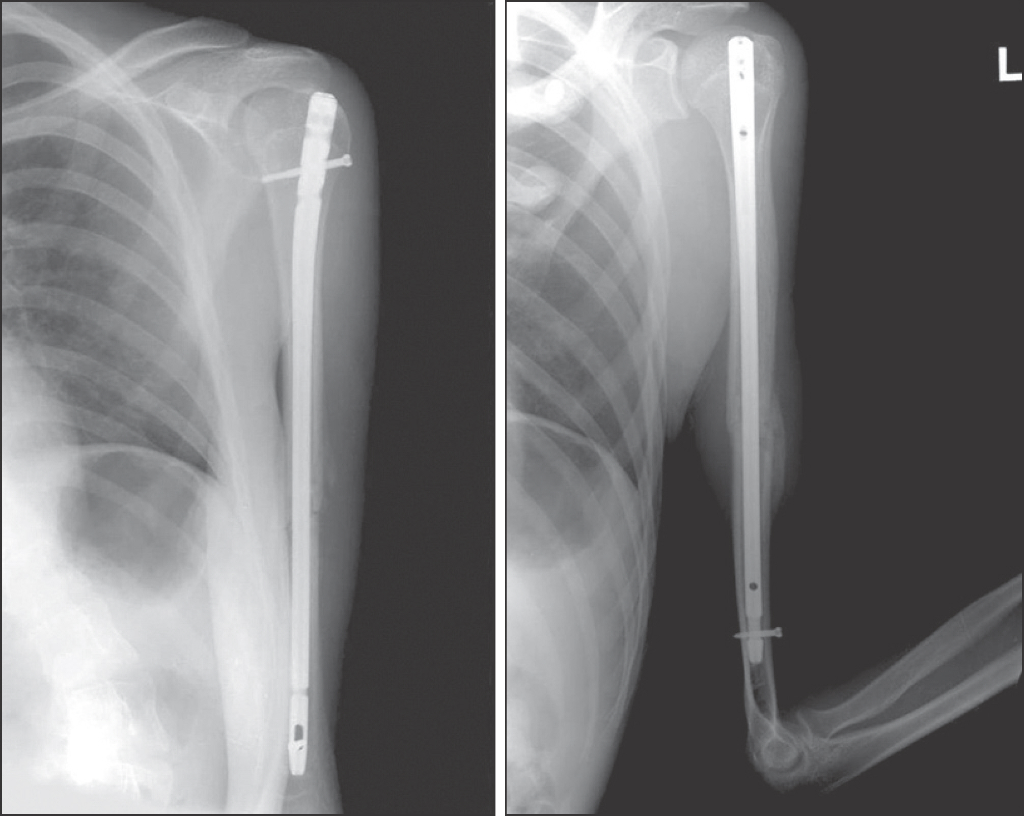
Figure 4. Radiographs 6 months after the operation show radiological bone union.
Reference
-
References
1. Papagelopoulos PJ, Galanis EC, Sim FH, Unni KK. Clinicopathologic features, diagnosis, and treatment of osteoblastoma. Orthopedics. 1999; 22:244–7.
Article2. Frassica FJ, Waltrip RL, Sponseller PD, Ma LD, McCarthy EF Jr. Clinicopathologic features and treatment of osteoid osteoma and osteoblastoma in children and adolescents. Orthop Clin North Am. 1996; 27:559–74.
Article3. Greenspan A. Benign bone-forming lesions: osteoma, osteoid osteoma, and osteoblastoma. Clinical, imaging, pathologic, and differential considerations. Skeletal Radiol. 1993; 22:485–500.
Article4. Marsh BW, Bonfiglio M, Brady LP, Enneking WF. Benign osteoblastoma: range of manifestations. J Bone Joint Surg Am. 1975; 57:1–9.5. Kirwan EO, Hutton PA, Pozo JL, Ransford AO. Osteoid osteoma and benign osteoblastoma of the spine. Clinical presentation and treatment. J Bone Joint Surg [Br]. 1984; 66:21–6.
Article6. Lichtenstein L. Benign osteoblastoma; a category of osteoid-and bone-forming tumors other than classical osteoid osteoma, which may be mistaken for giantcell tumor or osteogenic sarcoma. Cancer. 1956; 9:1044–52.7. Kroon HM, Schurmans J. Osteoblastoma: clinical and radiologic findings in 98 new cases. Radiology. 1990; 175:783–90.
Article8. Golant A, Lou JE, Erol B, Gaynor JW, Low DW, Dormans JP. Pediatric osteoblastoma of the sternum: a new surgical technique for reconstruction after removal: case report and review of the literature. J Pediatr Orthop. 2004; 24:319–22.9. Saglik Y, Atalar H, Yildiz Y, Basarir K, Gunay C. Surgical treatment of osteoblastoma: a report of 20 cases. Acta Orthop Belg. 2007; 73:747–53.
- Full Text Links
-
- Actions
-
Cited
- CITED
-
- Close
- Share
-
- Similar articles
-
- Malignant Osteoblastoma: A Case Report
- Humerus Shaft Fracture in a Wakeboarder
- A Comminuted Spiral Fracture with Butterfly Fragment of Distal Humerus by Arm Wrestling: A Case Report
- Medial Transposition of Radial Nerve in Distal Humerus Shaft Fracture: A Report of Six Cases
- The Comparative Study of Treatment between the IM nailing and the Plate fuation of the Humerus Shaft Fracture
