J Korean Soc Radiol.
2011 Jan;64(1):17-24. 10.3348/jksr.2011.64.1.17.
Computed Tomography Features of Follicular Thyroid Carcinoma: Comparison with Papillary Thyroid Carcinoma
- Affiliations
-
- 1Department of Radiology, Kangbuk Samsung Hospital, Sungkyunkwan University School of Medicine, Korea. yoonchoi99@yahoo.com
- KMID: 1443577
- DOI: http://doi.org/10.3348/jksr.2011.64.1.17
Abstract
- PURPOSE
The purpose of this study is to analyze the computed tomography (CT) features of follicular thyroid carcinoma (FTC) as compared to those of papillary thyroid carcinoma (PTC) to identify the characteristic imaging features of FTC.
MATERIALS AND METHODS
The following CT features were analyzed to compare FTCs (n = 17) and PTCs (n = 27): size, shape, margin, internal composition, calcification, enhancement homogeniety, extrathyroidal extension, lymph node (LN) metastasis and the density and degree of enhancement (qualitatively and quantitatively).
RESULTS
There were no significant differences between the patients with FTCs and those with PTCs with respect to age and gender, and the internal composition, calcification and enhancement homogeneity of the tumor. However, the FTCs tend to show a larger size (32 mm vs. 15 mm; respectively, p < 0.001), a round to oval shape (52.9% vs. 14.8% respectively, p = 0.001), a well-defined smooth margin (58.8% vs. 14.8% respectively, p = 0.009), less extrathyroidal extension (11.8% vs. 51.9% respectively, p = 0.007) and less LN metastasis (0% vs. 29.6% respectively, p = 0.016) compared to that of the PTCs. Furthermore, the FTCs showed significantly stronger enhancement in the early phase (146.4+/-42.4 vs. 98.5+/-38.2 respectively, p < 0.001) and a greater decrement of the late enhancement (-44.6+/-25.2 vs. -18.7+/-27.9 respectively, p = 0.003) compared to that of the PTCs.
CONCLUSION
FTCs had a tendency to be observed on neck CT as well-defined, smooth, round to oval nodules with strong early enhancement and a definite decrement of late enhancement as compared to the PTCs.
MeSH Terms
Figure
-
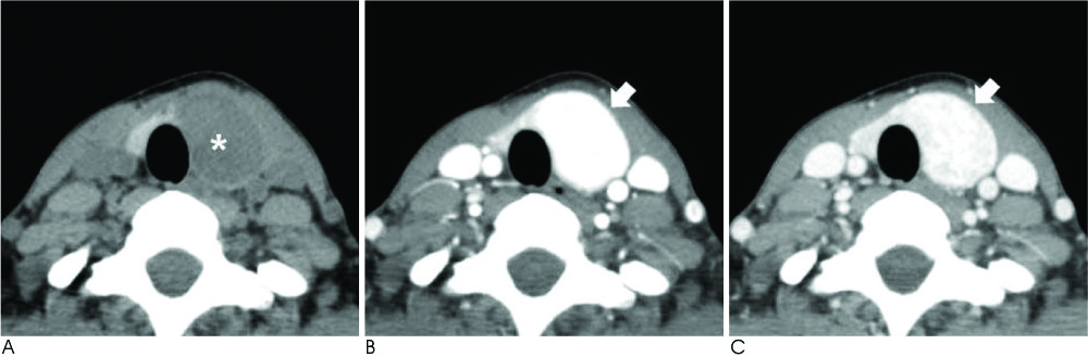
Fig. 1 Follicular thyroid carcinoma proven by surgical resection On the unenhanced CT (A), a well-defined smooth round to oval hypodense mass (*) is seen in the left lobe of the thyroid gland. On the early (B) and delayed phases (C), the mass (arrowed) shows strong enhancement relative to the thyroid parenchyma.
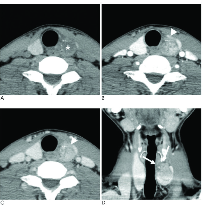
Fig. 2 Papillary thyroid carcinoma proven by surgical resection On the unenhanced CT (A), an ill-defined or irregular taller than wide hypodense mass (*) is seen in the left lobe of the thyroid gland. In the early (B) and delayed phases (C), the mass (arrowhead) shows less enhancement relative to the thyroid parenchyma. On the coronary MPR image (D), the mass shows definite extrathyroidal extension (arrows).
Reference
-
1. Widder S, Guggisberg K, Khalil M, Pasieka JL. A pathologic re-review of follicular thyroid neoplasms: the impact of changing the threshold for the diagnosis of the follicular variant of papillary thyroid carcinoma. Surgery. 2008; 144:80–85.2. Lin JD, Chao TC. Follicular thyroid carcinoma: from diagnosis to treatment. Endocr J. 2006; 53:441–448.3. Lo CY, Chan WF, Lam KY, Wan KY. Follicular thyroid carcinoma: the role of histology and staging systems in predicting survival. Ann Surg. 2005; 242:708–715.4. Albores-Saavedra J, Henson DE, Glazer E, Schwartz AM. Changing patterns in the incidence and survival of thyroid cancer with follicular phenotype--papillary, follicular, and anaplastic: a morphological and epidemiological study. Endocr Pathol. 2007; 18:1–7.5. Jeh SK, Jung SL, Kim BS, Lee YS. Evaluating the degree of conformity of papillary carcinoma and follicular carcinoma to the reported ultrasonographic findings of malignant thyroid tumor. Korean J Radiol. 2007; 8:192–197.6. Son KR, Na DG, Chang KH. Diagnostic value of CT for the detection of cervical lymph node metastases in papillary thyroid carcinoma. J Korean Soc Radiol. 2009; 60:383–389.7. AACE/AME Task Force on Thyroid Nodules. American Association of Clinical Endocrinologists and Associazione Medici Endocrinologi medical guidelines for clinical practice for the diagnosis and management of thyroid nodules. Endocr Pract. 2006; 12:63–102.8. Carling T, Udelsman R. Follicular neoplasms of the thyroid: what to recommend. Thyroid. 2005; 15:583–587.9. Ponikiewska D, Szczesniak-Klusek B, Stobiecka E, Jaworska M, Lange D. Oxyphilic and follicular thyroid tumors: the correlation between the cytopathologic diagnosis and the histopathologic examination. Endokrynol Pol. 2006; 57:Suppl A. 7–11.10. Frates MC, Benson CB, Doubilet PM, Cibas ES, Marqusee E. Can color Doppler sonography aid in the prediction of malignancy of thyroid nodules. J Ultrasound Med. 2003; 22:127–131. quiz 132-124.11. Lee B, Cook G, John L, Harrington K, Nutting C. Follicular thyroid carcinoma metastasis to the esophagus detected by 18FDG PET/CT. Thyroid. 2008; 18:267–271.12. Talbot JN, Montravers F, Younsi N, Zanotti-Fregonara P, Grahek D, Kerrou K, et al. PET in thyroid cancers. Presse Med. 2006; 35:1377–1385.13. Smith RB, Robinson RA, Hoffman HT, Graham MM. Preoperative FDG-PET imaging to assess the malignant potential of follicular neoplasms of the thyroid. Otolaryngol Head Neck Surg. 2008; 138:101–106.14. Choi YJ, Yun JS, Kim DH. Clinical and ultrasound features of cytology diagnosed follicular neoplasm. Endocr J. 2009; 56:383–389.15. Sillery JC, Reading CC, Charboneau JW, Henrichsen TL, Hay ID, Mandrekar JN. Thyroid follicular carcinoma: sonographic features of 50 cases. AJR Am J Roentgenol. 2010; 194:44–54.16. Yoon DY, Chang SK, Choi CS, Yun EJ, Seo YL, Nam ES, et al. The prevalence and significance of incidental thyroid nodules identified on computed tomography. J Comput Assist Tomogr. 2008; 32:810–815.17. Shetty SK, Maher MM, Hahn PF, Halpern EF, Aquino SL. Significance of incidental thyroid lesions detected on CT: correlation among CT, sonography, and pathology. AJR Am J Roentgenol. 2006; 187:1349–1356.18. Lang BH, Lo CY, Chan WF, Lam KY, Wan KY. Staging systems for follicular thyroid carcinoma: application to 171 consecutive patients treated in a tertiary referral centre. Endocr Relat Cancer. 2007; 14:29–42.19. Jung JH, Hwang GH, Park HY, Lee YH. Role of Ultrasonography in differential diagnosis of thyroid nodules. J Korean Surg Soc. 2006; 70:349–356.20. Fukunari N, Nagahama M, Sugino K, Mimura T, Ito K. Clinical evaluation of color Doppler imaging for the differential diagnosis of thyroid follicular lesions. World J Surg. 2004; 28:1261–1265.21. Miyakawa M, Onoda N, Etoh M, Fukuda I, Takano K, Okamoto T, et al. Diagnosis of thyroid follicular carcinoma by the vascular pattern and velocimetric parameters using high resolution pulsed and power Doppler ultrasonography. Endocr J. 2005; 52:207–212.22. Paramo JC, Mesko T. Age, tumor size, and in-office ultrasonography are predictive parameters of malignancy in follicular neoplasms of the thyroid. Endocr Pract. 2008; 14:447–451.23. Kondo T, Ezzat S, Asa SL. Pathogenetic mechanisms in thyroid follicular-cell neoplasia. Nat Rev Cancer. 2006; 6:292–306.24. Oertel YC. Follicular lesions of the thyroid gland: from hyperplasia to neoplasia. Endocr Pract. 2008; 14:251–252.25. Sillery JC, Reading CC, Charboneau JW, Henrichsen TL, Hay ID, Mandrekar JN. Thyroid follicular carcinoma: sonographic features of 50 cases. AJR Am J Roentgenol. 2010; 194:44–54.26. Gharib H, Papini E, Valcavi R, Baskin HJ, Crescenzi A, Dottorini ME, et al. American Association of Clinical Endocrinologists and Associazione Medici Endocrinologi medical guidelines for clinical practice for the diagnosis and management of thyroid nodules. Endocr Pract. 2006; 12:63–102.
- Full Text Links
-
- Actions
-
Cited
- CITED
-
- Close
- Share
-
- Similar articles
-
- A Case of Mixed Follicular-Papillary Thyroid Carcinoma
- Concurrent Medullay and Papillary Carcinoma of the Thyroid
- Oxyphilic Papillary Carcinoma of the Thyroid in Fine Needle Aspiration
- Demonstration of TCM-9 Monoclonal Antibody in Follicular Neoplasm of Thyroid
- Tremendous Skull Metastasis from Follicular Thyroid Carcinoma: Case Reort
