Multidetector CT Findings of Retroarterial Reversed Intestinal Rotation in an Adult: A Case Report
- Affiliations
-
- 1Department of Radiology, Uijeongbu St. Mary's Hospital, College of Medicine, The Catholic University of Korea, Korea. radlsl@catholic.ac.kr
- 2Department of Internal Medicine, Uijeongbu St. Mary's Hospital, College of Medicine, The Catholic University of Korea, Korea.
- KMID: 1443540
- DOI: http://doi.org/10.3348/jksr.2011.64.4.365
Abstract
- Intestinal malrotation is an uncommon cause of abdominal pain in adults. More so, reversed intestinal rotation is one of the least common malrotation anomalies. A few cases of reversed intestinal rotation have been reported only in surgical and internal medicine literatures. To the best of our knowledge, MDCT findings concerning reversed intestinal rotation had never been reported. We present a 38-year-old man with retroarterial reversed intestinal rotation with MDCT findings.
Figure
-
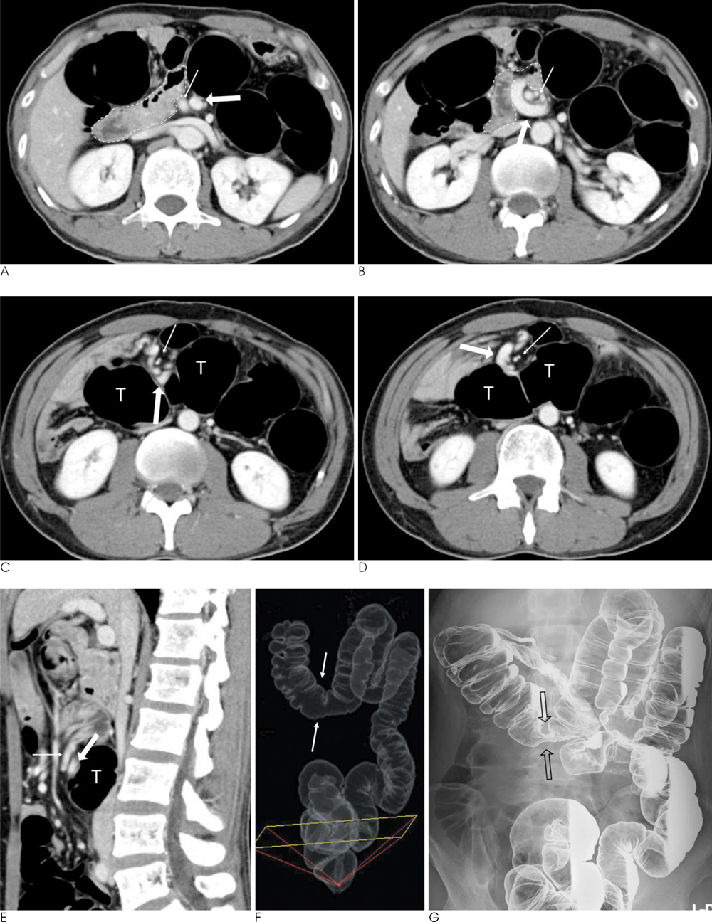
Fig. 1 A-D. Serial axial images of a contrast-enhanced CT of a 38-year-old man with reversed rotation. SMV (thick arrow) lies to the left of SMA (thin arrow) in the first image (A), and SMV wraps around SMA clockwise on consecutive images (B-D). It is noted that the duodenum (dotted lines in A and B) cross from the right to left anterior to SMA and SMV. Images at the level of lower pole of the kidneys (Cand D) demonstrate that the distended transverse colon (T) is sandwiched between the superior mesenteric vessels and the aorta. E-G. The sagittal (F) reformatted image more clearly depicted the relationship between the superior mesenteric vessels (thin and thick arrows) and the transverse colon (T). A tissue transition projection (F) from CT colonoscopy discloses focal narrowing of transverse colon (open arrows) and the abnormal location of the cecum. The overhead image after double-contrast barium enema (G) is exactly coincidental with the image of CT colonoscopy, which shows focal extrinsic compression of the transverse colon and resultant narrowing of the involved segment (open arrows). The cecum, appendix, and ileocecal valve are located in the right upper quadrant of the abdomen.
Reference
-
1. Aldridge RT. Intestinal malrotation in the adult. N Z Med J. 1996; 60:420–423.2. O'Connell P, Lynch G. Reversed intestinal rotation associated with anomalous mesenteric venous drainage. Dis Colon Rectum. 1990; 33:883–885.3. Berardi RS. Anomalies of midgut rotation in the adult. Surg Gynecol Obstet. 1980; 151:113–124.4. Lilleyman A, Levy RD, Sillar R. Reversed intestinal rotation: report of two cases and review of the published reports. ANZ J Surg. 2006; 76:947–949.5. Cunningham T, Hartman G, Bulas DI. CT findings in prearterial reversed midgut rotation. Pediatr Radiol. 1994; 24:537–538.6. Estrada RL, Gurd FN. Surgical correction of reversed rotation of the midgut loop. Surg Gynecol Obstet. 1962; 114:707–711.7. Nehra D, Zeineh M, Rodriguez F, Dutta S. Double reverse intestinal malrotation: a novel rotational anomaly and its surgical correction. J Pediatr Surg. 2007; 42:578–581.8. Shimanuki Y, Aihara T, Takano H, Moritani T, Oguma E, Kuroki H, et al. Clockwise whirlpool sign at color Doppler US: an objective and definite sign of midgut volvulus. Radiology. 1996; 199:261–264.9. Clark P, Ruess L. Counterclockwise barber-pole sign on CT: SMA/SMV variance without midgut malrotation. Pediatr Radiol. 2005; 35:1125–1127.
- Full Text Links
-
- Actions
-
Cited
- CITED
-
- Close
- Share
-
- Similar articles
-
- Radiologic Findings of Reversed Intestinal Rotation in Adults: 3 Cases Report
- Multidetector Computed Tomography Findings of Mesenteroaxial Gastric Volvulus Combined with Torsion of Wandering Spleen: A Case Report and Literature Review
- Unilateral Pulmonary Vein Atresia: A Case Report
- Computed Tomographic Findings of Anomalies of Intestinal Rotation in Adult Patients
- A case of intestinal non-rotation incidentally detected on DISIDA scan
