Treatment of Spinal Osseous Metastasis with Combined Percutaneous Radiofrequency Ablation and Vertebroplasty
- Affiliations
-
- 1Department of Radiology, Korea University Guro Hospital, Korea University College of Medicine, Seoul, Korea. hongsj@korea.ac.kr
- 2Department of Neurosurgery, Korea University Guro Hospital, Korea University College of Medicine, Seoul, Korea.
- KMID: 1443536
- DOI: http://doi.org/10.3348/jksr.2011.64.4.333
Abstract
- Recent introduction of image-guided percutaneous methods to treat unresectable bone tumors including metastases that do not respond to conventional radiotherapy or chemotherapy has proven to be effective. Here we present three successfully treated cases of metastatic bone lesions: two cases of malignant bone metastases in the lumbar spine and one in the sacral bone, using combined percutaneous radiofrequency ablation and percutaneous vertebroplasty/cementoplasty. A brief review of literature is also included.
Figure
-
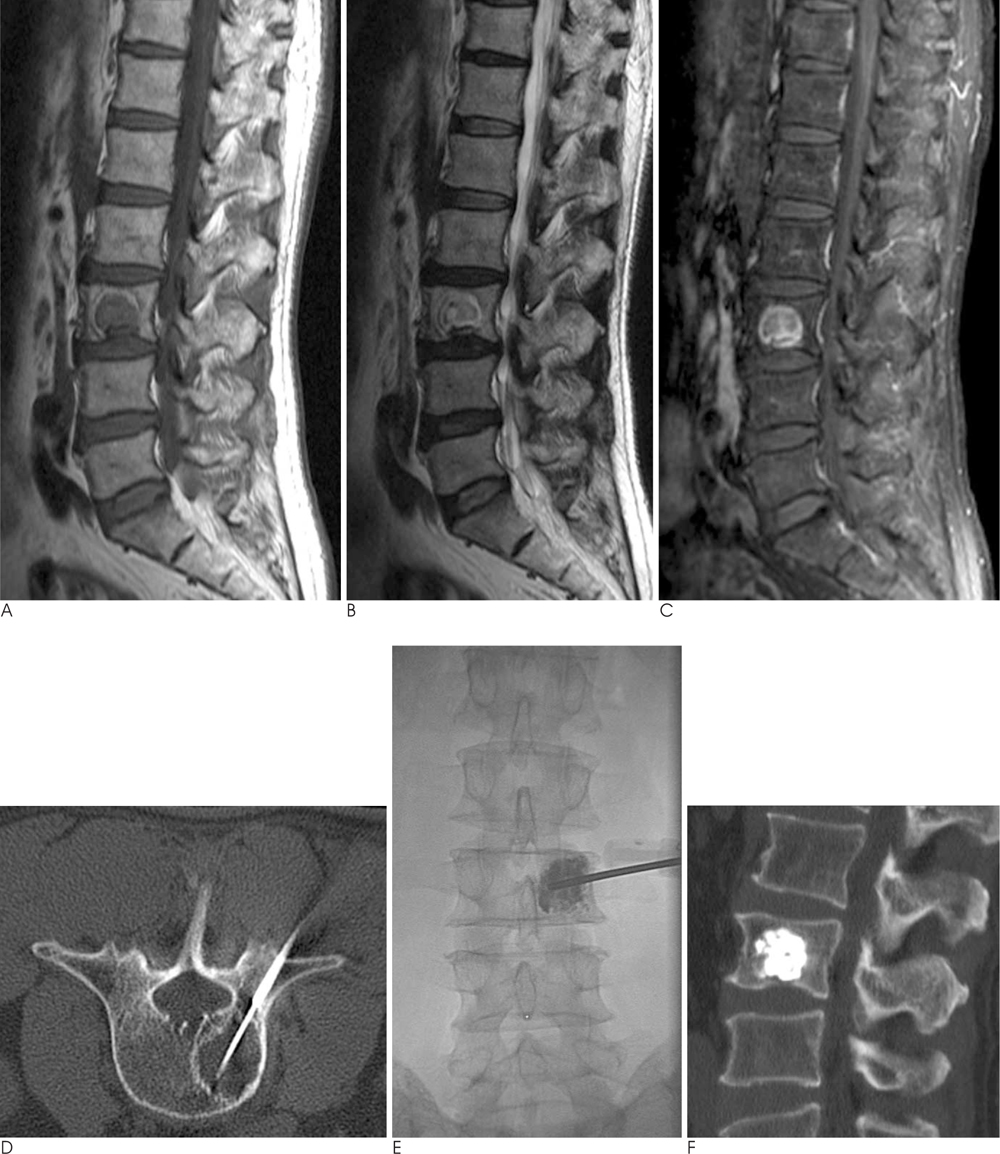
Fig. 1 RFA and combined percutaneous vertebroplasty of spinal metastasis in a 59-year-old man with hepatocelluar carcinoma. Sagittal T1 (A), T2 (B), and post-contrast fat suppressed T1 (C) - weighted images of the lumbar spine show a round mass lesion in the L3 body with T1 low, T2 heterogeneous high signal intensity, diffuse and prominent peripheral enhancement, representing a metastatic lesion. Axial CT scan during RFA (D) demonstrates the inserted RFA needle with a 3 cm active tip in the center of the lesion through the 11-gauge vertebroplasty cannula. Bone cement is well packed in the center of the metastatic lesion on the immediate prone AP view (E) and sagittal CT scan (F) after combined vertebroplasty.
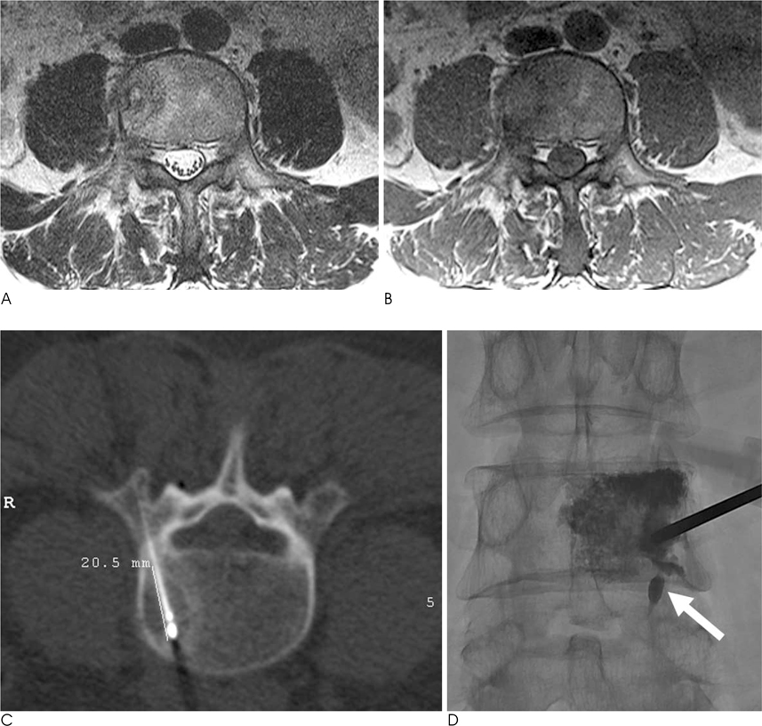
Fig. 2 A 49-year-old man with hepatocelluar carcinoma and lumbar metastasis treated with RFA and combined percutaneous vertebroplasty. Axial T2 (A), T1 (B) - MR images show a heterogeneous T2 high, T1 low signal intensity mass with surrounding bone edema in the right side of the L4 body. Axial CT scan during RFA (C) demonstrates an inserted RFA needle with a 2 cm active tip in the center of the lesion. Immediate prone AP radiograph (D) shows injected cement in the lesion and surrounding bone marrow of the L4 body. Small amount of intradiscal cement leakage is seen (arrow).
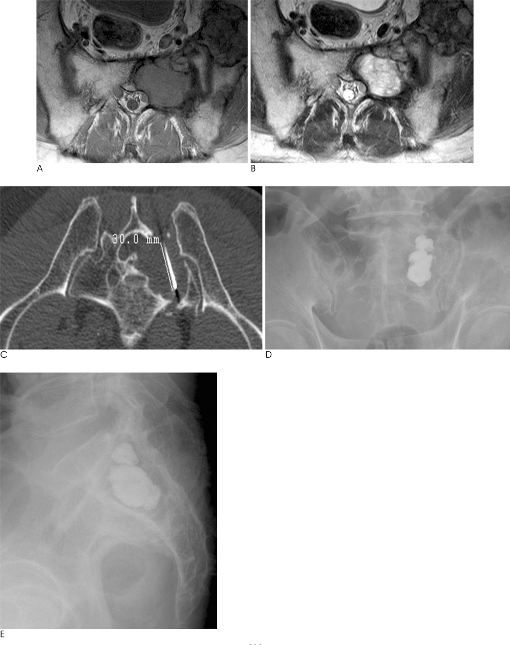
Fig. 3 Metastatic renal cell carcinoma involving the left sacrum in a 67-year-old man. Axial T1- (A), T2- (B) weighted MR images show an inhomogeneous T1 low, T2 high signal mass in left sacral ala with bone expansion. Axial CT scan during RFA (C) shows the needle inserted in the center of the left sacral lesion. Frontal (D) and lateral (E) radiographs taken after RFA and cementoplasty show well confinement of polymethylmetacrylate within the sacral mass lesion.
Reference
-
1. Callstrom MR, Charboneau JW, Goetz MP, Rubin J, Atwell TD, Farrell MA, et al. Image-guided ablation of painful metastatic bone tumors: a new and effective approach to a difficult problem. Skeletal Radiol. 2006; 35:1–15.2. Dupuy DE, Hong R, Oliver B, Goldberg SN. Radiofrequency ablation of spinal tumors: temperature distribution in the spinal canal. AJR Am J Roentgenol. 2000; 175:1263–1266.3. Goetz MP, Callstrom MR, Charboneau JW, Farrell MA, Maus TP, Welch TJ, et al. Percutaneous image-guided radiofrequency ablation of painful metastases involving bone: a multicenter study. J Clin Oncol. 2004; 22:300–306.4. Hoffmann RT, Jakobs TF, Trumm C, Weber C, Helmberger TK, Reiser MF. Radiofrequency ablation in combination with osteoplasty in the treatment of painful metastatic bone disease. J Vasc Interv Radiol. 2008; 19:419–425.5. Gronemeyer DH, Schirp S, Gevargez A. Image-guided radiofrequency ablation of spinal tumors: preliminary experience with an expandable array electrode. Cancer J. 2002; 8:33–39.6. Toyota N, Naito A, Kakizawa H, Hieda M, Hirai N, Tachikake T, et al. Radiofrequency ablation therapy combined with cementoplasty for painful bone metastases: initial experience. Cardiovasc Intervent Radiol. 2005; 28:578–583.7. Schaefer O, Lohrmann C, Herling M, Uhrmeister P, Langer M. Combined radiofrequency thermal ablation and percutaneous cementoplasty treatment of a pathologic fracture. J Vasc Interv Radiol. 2002; 13:1047–1050.8. Nakatsuka A, Yamakado K, Maeda M, Yasuda M, Akeboshi M, Takaki H, et al. Radiofrequency ablation combined with bone cement injection for the treatment of bone malignancies. J Vasc Interv Radiol. 2004; 15:707–712.9. van der Linden E, Kroft LJ, Dijkstra PD. Treatment of vertebral tumor with posterior wall defect using image-guided radiofrequency ablation combined with vertebroplasty: preliminary results in 12 patients. J Vasc Interv Radiol. 2007; 18:741–747.
- Full Text Links
-
- Actions
-
Cited
- CITED
-
- Close
- Share
-
- Similar articles
-
- Tageted bipolar radiofrequency decompression with vertebroplasty for intractable radicular pain due to spinal metastasis: a case report
- Two Cases of Percutaneous Vertebroplasty for Spinal Metastatic Cancer: A case report
- Percutaneous Vertebroplasty in Spinal Metastasis and Myeloma:25 Cases Experience
- Lumbar Root Injury by the Leakage of Bone Cement after the Percutaneous Vertebroplasty: A case report
- The Comparision of the Radiofrequency Ablasion Therapy with Vertebroplasty and Radiotherapy in Metastatic Spine Tumor
