Inflammatory Pseudotumor of the Pituitary Gland Mimicking a Pituitary Adenoma: A Case Report
- Affiliations
-
- 1Department of Radiology, Eulji University Hospital, Daejeon, Korea. midosyu@eulji.ac.kr
- 2Department of Neurosurgery, Eulji University Hospital, Daejeon, Korea.
- 3Department of Pathology, Eulji University Hospital, Daejeon, Korea.
- KMID: 1443531
- DOI: http://doi.org/10.3348/jksr.2011.64.4.303
Abstract
- A 38-year-old man was admitted to our hospital with diplopia. The patient had a relatively well-defined pituitary mass with high cellularity as well as weaker enhancement on imaging modalities including computed tomography (CT) and magnetic resonance imaging (MRI), than a typical pituitary adenoma. The distinction between a pseudotumor and an invasive neoplasm is very difficult before biopsy. In this case report, we discuss the characteristic imaging features of a fibrosing inflammatory pseudotumor of the pituitary gland.
MeSH Terms
Figure
-
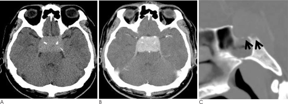
Fig. 1 A 38-year-old man presenting with ophthalmoplegia and diplopia that aggravated the left lateral gaze. A. Pre-contrast CT image showing a well defined, homogeneously high attenuated lesion in the sella and parasellar regions. B. Postcontrast CT image shows homogeneously well enhanced lesion with encasement of both cavernous ICA. C. Sagittal bone algorithm CT image showing permeative bone erosion of the sellar floor and dorsum sella (arrows).
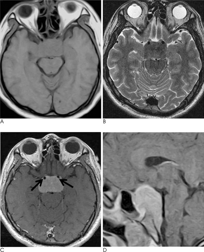
Fig. 2 A. T1-weighed axial MR image showing a well defined iso-signal intensity lesion, involving the sellar and parasellar region. B. T2-weighted axial MR image showing a well defined low signal intensity lesion, involving the sellar and parasellar region. C. Contrast-enhanced T1-weighted axial MR image showing homogenous enhancement of the lesion with bilateral involvement of the cavernous sinuses associated with encasement of the both cavernous ICA (arrows). D. Contrast-enhanced T1-weighted sagittal MR image shows the caudal portion of the lesion extending to the retrosellar region. Note that minimal suprasellar bulging contour of the mass was evident despite the involvement of both cavernous sinuses.

Fig. 3 A. Left ICA angiogram showing tumor staining in the venous phase, similar to a meningioma, but with a weaker degree of demarcation than staining of the meningioma. B. Left ICA angiogram showing focal severe stenosis of the Lt. distal cavernous ICA in the arterial phase. C. Photomicrograph of the inflammatory pseudotumor (inflammatory myofibroblastic tumor) of the pituitary TSA specimen showing underlying collagenous dense fibrous tissue (asterisk) which was infiltrated by prominent inflammatory infiltrates (mainly plasma cells, lymphocytes, and histiocytes) with a Russel body (small, pink colored, spherical intracytoplasmic hyaline body, arrows) (H-E stain, magnification, × 400).
Reference
-
1. Lee JH, Kim KJ, Chung SW, Choi YC, Lee Ah. A case report of inflammatory pseudotumor involving the clivus : CT and MR findings. Korean J Radiol. 2001; 2:231–234.2. Al-Shraim M, Syro LV, Kovacs K, Estrada H, Uribe H, Al-Gahtany M. Inflammatory pseudotumor of the pituitary gland: case report. Surg Neurol. 2004; 62:264–267.3. Hansen I, Petrossians P, Thiry A, Flandroy P, Gaillard RC, Lovacs K, et al. Extensive inflammatory pseudotumor of the pituitary. J Clin Endocrinol Metab. 2001; 86:4603–4610.4. Murakami K, Muraishi K, Ikeda H, Yoshimoto T. Plasma cell granuloma of the pituitary gland. Surg Neurol. 2001; 56:247–251.5. Choi SY, Yu IK, Han MH, Lee BH, Song CJ, Kim KS. Fibrosing inflammatory pseudotumor of the nasopharynx: MR features and histopathologic correlation. Euro J Radiol. 2009; 72:274–277.6. Mombaerts I, Goldschmeding R, Schlingemann RO, Koornneef L. What is orbital pseudotumor? Surv Ophthalmol. 1996; 41:66–78.7. Swain RS, Tihan T, Horvai AE, Di Vizio D, Loda M, Burger PC, et al. Inflammatory myofibroblastic tumor of the central nervous system and its relationship to inflammatory pseudotumor. Hum Pathol. 2008; 39:410–419.8. Han MH, Chi JG, Kim MS, Chang KH, Kim KH, Yeon KM, et al. Fibrosing inflammatory pseudotumors involving the skull base : MR and CT manifestations with histopathologic comparison. AJNR Am J Neuroradiol. 1996; 17:515–521.9. Seider MJ, Cleary KR, Van Tassel P, Alexanian R, Schantz SP, Frias A, et al. Plasma cell granuloma of the nasal cavity treated by radiation therapy. Cancer. 1991; 67:929–932.10. Char DH, Miller T. Orbital pseudotumor. Fine needle aspiration biopsy and response to therapy. Ophthalmology. 1993; 100:1702–1170.
- Full Text Links
-
- Actions
-
Cited
- CITED
-
- Close
- Share
-
- Similar articles
-
- Pituitary Apoplexy due to Pituitary Adenoma Infarction
- Two Cases of Pituitary Hyperplasia Secondary to Primary Hypothyroidism Mimicking Pituitary Tumor
- A Case of Acromegaly Caused by Mixed Gangliocytoma-Adenoma of the Pituitary Gland
- Computed tomography of pituitary apoplexy: report of 2 cases
- Metastasis of Hepatocellular Carcinoma to Pituitary Adenoma: A Case Report
