J Korean Soc Radiol.
2011 Jul;65(1):9-12. 10.3348/jksr.2011.65.1.9.
Pituitary Abscess Presenting a Pituitary Tumor: A Case Report
- Affiliations
-
- 1Department of Radiology, Gangneung Asan Hospital, College of Medicine, University of Ulsan, Gangneung, Korea. neurorad@lycos.co.kr
- KMID: 1443486
- DOI: http://doi.org/10.3348/jksr.2011.65.1.9
Abstract
- A pituitary abscess is a rare disease accounting for approximately 1% or less of all pituitary lesions. We report a pathologically proven pituitary abscess mimicking the pituitary tumor.
Figure
-
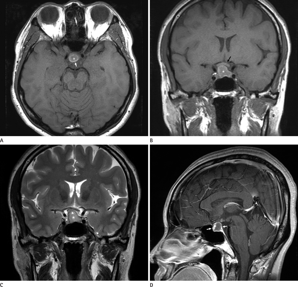
Fig. 1 A 39-year-old female with a pituitary abscess. A, B. Axial (A) and coronal (B) T1-weighted (500/10) images show a huge mass in the sellar and suprasellar area with mild compression of the optic chiasm (black arrow). The mass shows hyposignal intensity (white asterisk) with round hyperintense rim (white arrow). C. Coronal T2-weighted image (3000/80) shows high signal intensity mass (white asterisk) with a thin hypointense rim (white arrowheads). D. Sagittal contrast enhanced T1-weighted (500/10) image shows a rim-enhancing mass (white arrows).
Reference
-
1. Vates GE, Berger MS, Wilson CB. Diagnosis and management of pituitary abscess: a review of twenty-four cases. J Neurosurg. 2001; 95:233–241.2. Dutta P, Bhansali A, Singh P, Kotwal N, Pathak A, Kumar Y. Pituitary abscess: report of four cases and review of literature. Pituitary. 2006; 9:267–273.3. Kabuto M, Kubota T, Kobayashi H, Takeuchi H, Kubota T, Nakagawa T, et al. MR imaging and CT of pituitary abscess: case report and review. Neurol Res. 1996; 18:495–498.4. Wolansky LJ, Gallagher JD, Heary RF, Malantic GP, Dasmahapatra A, Shaderowfsky PD, et al. I of pituitary abscess: two cases and review of the literature. Neuroradiology. 1997; 39:499–503.5. Sabbah P, Bonardel G, Herve R, Marjou F, Hor F, Pharaboz C, et al. CT and MRI findings in primitive pituitary abscess: a case report and review of literature. J Neuroradiol. 1999; 26:196–199.6. Shuster A, Gunnarsson T, Sommer D, Miller E. Pituitary abscess: an unexpected diagnosis. Pediatr Radiol. 2010; 40:219–222.7. Ovali GY, Tarhan S, Şentürk H, Bayindir P, Pabuşçu Y. Pituitary abscess simulating macroadenoma. Turk J Med Sci. 2004; 34:337–339.8. Lemoncito MV, Lantion-Ang FL. Pituitary abscess mimicking pituitary adenoma: a review of three cases seen at the Philippine General Hospital from 2004-2007. Phil J Int Med. 2009; 47:143–150.9. Zimmerman RD, Weingarten K. Neuroimaging of cerebral abscess. Neuroimaging Clin N Am. 1991; 1:1–16.10. Haimes AB, Zimmerman RD, Morgello S, Weingarten K, Becker RD, Jennis R, et al. MR imaging of brain abscesses. AJR Am J Roentgenol. 1989; 152:1073–1085.
