Focal Fat Deposition Developed in the Segment IV of the Liver Following Gastrectomy Mimicking a Hepatic Metastasis: Two Case Reports
- Affiliations
-
- 1Department of Radiology and Research Institute of Radiological Science, Severance Hospital, Yonsei University College of Medicine, Seoul, Korea. jslim1@yuhs.ac
- 2Department of Surgery, Severance Hospital, Yonsei University College of Medicine, Seoul, Korea.
- KMID: 1439537
- DOI: http://doi.org/10.3348/jksr.2012.67.4.257
Abstract
- We present two cases of focal fat deposition developed at the posterior area of the segment IV in the liver, following gastrectomy in patients with gastric cancer. There was no focal lesion in this area of the liver at preoperative computed tomography (CT) in both cases, and the aberrant right gastric vein (ARGV) was found on the retrospective review of this CT. After gastrectomy, a focal, low-attenuating lesion was developed in this area on a follow-up CT in both cases, which was confirmed as a focal fat deposition, by other imaging studies. In addition to its typical imaging findings, confirmation of the presence of the ARGV also supported this lesion to be a focal fat deposition. Furthermore, understanding of our cases may be of help to prevent us from unnecessary invasive procedures, such as liver biopsy.
Figure
-
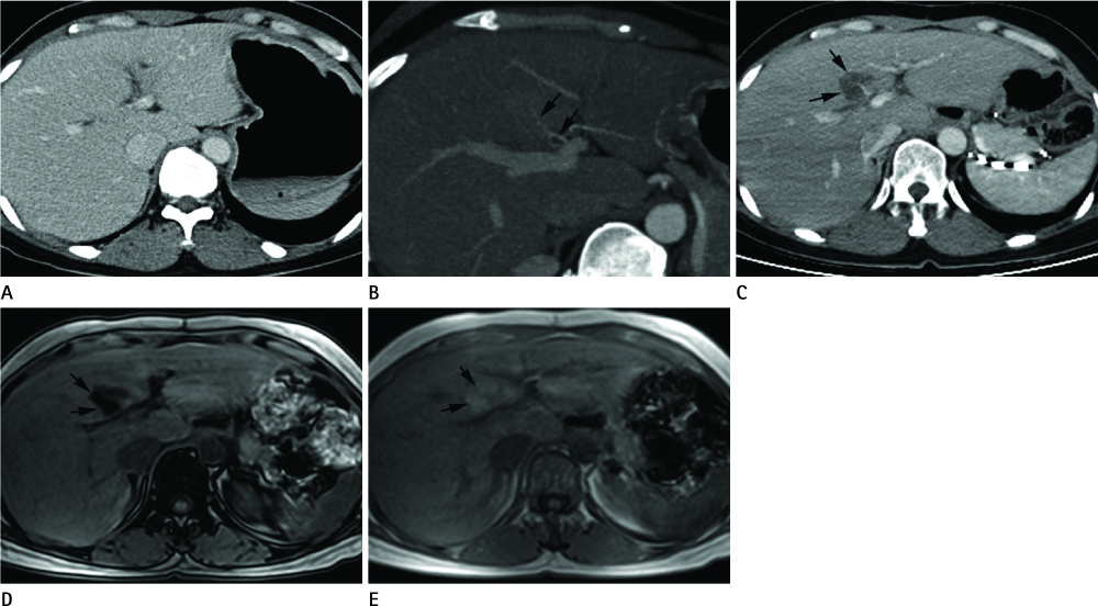
Fig. 1 A 44-year-old woman who underwent total gastrectomy due to advanced gastric cancer. A. Preoperative axial CT image obtained during the portal venous phase demonstrates no focal lesion in the segment IV of the liver. B. Preoperative oblique CT image using the maximum intensity projection technique demonstrates an enhancing vascular structure (arrows) at the posterior area of the segment IV, which was found to originate from the aberrant right gastric vein on the other maximum intensity projection images. C. Follow-up CT image obtained one year after gastrectomy demonstrates a 2.5-cm, low-attenuating lesion (arrows) newly developed in the segment IV. Note an enhancing vascular structure within this lesion. D, E. Chemical shift magnetic resonance images confirm that this lesion (arrows) is focal fat deposition by identifying signal drop of this lesion on the opposed phase image (D) in comparison with the in-phase image (E).

Fig. 2 A 69-year-old man who underwent subtotal gastrectomy due to advanced gastric cancer. A. Preoperative axial CT image obtained during the portal venous phase demonstrates no focal lesion in the segment IV of the liver. B. Preoperative coronal CT image using the maximum intensity projection technique demonstrates the ARGV (arrows) that drains into the posterior area of segment IV. The common hepatic artery is also visualized (arrowheads). C. Follow-up CT image obtained six months after gastrectomy demonstrates a 2.9-cm, low-attenuating lesion (arrows) newly developed in the segment IV. Note an enhancing vascular structure within this lesion. D. On the one-year follow-up CT image, the lesion becomes to be smaller and less distinct, due to partial recovery of attenuation at the posterior part of the lesion (arrows). Note.-ARGV = aberrant right gastric vein
Reference
-
1. Matsui O, Takahashi S, Kadoya M, Yoshikawa J, Gabata T, Takashima T, et al. Pseudolesion in segment IV of the liver at CT during arterial portography: correlation with aberrant gastric venous drainage. Radiology. 1994. 193:31–35.2. Matsui O, Kadoya M, Takahashi S, Yoshikawa J, Gabata T, Takashima T, et al. Focal sparing of segment IV in fatty livers shown by sonography and CT: correlation with aberrant gastric venous drainage. AJR Am J Roentgenol. 1995. 164:1137–1140.3. Yates CK, Streight RA. Focal fatty infiltration of the liver simulating metastatic disease. Radiology. 1986. 159:83–84.4. Yoshimitsu K, Honda H, Kuroiwa T, Irie H, Aibe H, Shinozaki K, et al. Unusual hemodynamics and pseudolesions of the non-cirrhotic liver at CT. Radiographics. 2001. 21 Spec No:S81–S96.5. Yamagami T, Arai Y, Takeuchi Y, Sueyoshi S, Inaba Y. Focal fatty change in segment IV of the liver occurring after gastrectomy. Br J Radiol. 1998. 71:888–891.6. Lee JW, Kim S, Kwack SW, Kim CW, Moon TY, Lee SH, et al. Hepatic capsular and subcapsular pathologic conditions: demonstration with CT and MR imaging. Radiographics. 2008. 28:1307–1323.7. Yoshimitsu K, Irie H, Kakihara D, Tajima T, Asayama Y, Hirakawa M, et al. Postgastrectomy development or accentuation of focal fatty change in segment IV of the liver: correlation with the presence of aberrant venous branches of the parabiliary venous plexus. J Clin Gastroenterol. 2007. 41:507–512.8. Sohn J, Siegelman E, Osiason A. Unusual patterns of hepatic steatosis caused by the local effect of insulin revealed on chemical shift MR imaging. AJR Am J Roentgenol. 2001. 176:471–474.9. Mitchell DG, Kim I, Chang TS, Vinitski S, Consigny PM, Saponaro SA, et al. Fatty liver. Chemical shift phase-difference and suppression magnetic resonance imaging techniques in animals, phantoms, and humans. Invest Radiol. 1991. 26:1041–1052.
- Full Text Links
-
- Actions
-
Cited
- CITED
-
- Close
- Share
-
- Similar articles
-
- F-18 FDG Uptake in an Eosinophilic Liver Abscess Mimicking Hepatic Metastasis on PET/CT Images
- Liver Metastasis Mimicking Cholangiocarcinoma from Colon Cancer
- CT Findings of Ciliated Hepatic Foregut Cyst Mimicking Metastasis: A Case Report
- Hepatocellular Adenoma and Focal Nodular Hyperplasia
- Focal Nodular Hyperplasis in Liver
