Gradual Progression of Intrapulmonary Lymph Nodes Associated with Usual Interstitial Pneumonia in Progressive Systemic Sclerosis on Chest Radiographs and CT
- Affiliations
-
- 1Department of Radiology, Bucheon St. Mary's Hospital, College of Medicine, The Catholic University of Korea, Bucheon, Korea. corvidae@daum.net
- 2Department of Radiology, Seoul St. Mary's Hospital, College of Medicine, The Catholic University of Korea, Seoul, Korea.
- 3Department of Internal Medicine, Bucheon St. Mary's Hospital, College of Medicine, The Catholic University of Korea, Bucheon, Korea.
- KMID: 1439533
- DOI: http://doi.org/10.3348/jksr.2012.67.4.241
Abstract
- A 40-year-old female visited the clinic for evaluation of Raynaud's phenomenon for a period of four years. The initial chest radiograph showed a fine reticular density and ground glass opacity with lower lobe predominance. These findings are consistent interstitial fibrosis. Additionally, high resolution CT showed multiple, small, coexisting nodular opacities, ranging from 3 to 7 mm in size in both lungs. These nodules grew up to 1.5 cm and showed moderate enhancement. Because of the rareness of intrapulmonary lymph node in patient of progressive systemic sclerosis, we couldn't exclude the possibility of malignancy. These nodules are turned out to be intrapulmonary lymph nodes on video-assisted thoracoscopic lung biopsy.
MeSH Terms
Figure
-
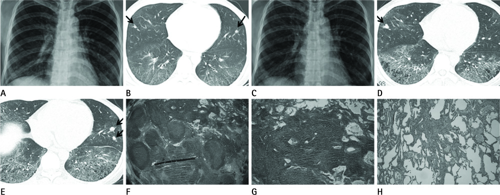
Fig. 1 A 40-year-old female with history of Raynaud's phenomenon. A. The initial chest radiograph shows diffuse fine reticular densities and ground-glass opacities in both lung fields with basal lung predominance. B. HRCT shows fine reticulonodular densities and diffuse ground-glass opacities in the right middle lobe, both lower lobes and left lingular segment. Findings are compatible with interstitial lung disease due to PSS. Nodules in the right middle lobe and left lingular segments (arrows) were overlooked at this point of time. C. Six years later, follow-up chest radiograph shows irregular reticulonodular opacities bilaterally. The lung volume was slightly decreased. D, E. Follow-up HRCT shows interval increase in size of the nodules in the right middle lobe (arrow) and left lingular segment (arrow). The nodules in the right middle lobe become lobulated. Nodules are found more than 1 cm away from the pleura. There are increased in the honeycomb cysts. F, G. Histologic examination of the nodules reveals a lymph node with lymphoid follicles in the lung parenchyma (Hematoxylin and Eosin stain, × 100). H. Lung tissue obtained by wedge resection of the right lower lobe shows interstitial pneumonia compatible with the usual interstitial pneumonia pattern, due to the moderate activity of PSS (Hematoxylin and Eosin stain, × 100). Note.-HRCT = high resolution CT, PSS = progressive systemic sclerosis
Reference
-
1. Arroliga AC, Podell DN, Matthay RA. Pulmonary manifestations of scleroderma. J Thorac Imaging. 1992. 7:30–45.2. Silver RM, Metcalf JF, Stanley JH, LeRoy EC. Interstitial lung disease in scleroderma. Analysis by bronchoalveolar lavage. Arthritis Rheum. 1984. 27:1254–1262.3. Harrison NK, Myers AR, Corrin B, Soosay G, Dewar A, Black CM, et al. Structural features of interstitial lung disease in systemic sclerosis. Am Rev Respir Dis. 1991. 144(3 Pt 1):706–713.4. Howling SJ, Hansell DM, Wells AU, Nicholson AG, Flint JD, Müller NL. Follicular bronchiolitis: thin-section CT and histologic findings. Radiology. 1999. 212:637–642.5. Szekanecz E, Szamosi S, Gergely L, Keszthelyi P, Szekanecz Z, Szucs G. Incidence of lymphoma in systemic sclerosis: a retrospective analysis of 218 Hungarian patients with systemic sclerosis. Clin Rheumatol. 2008. 27:1163–1166.6. Yoshitomi A, Sato A, Toyoshima M, Suganuma H, Imokawa S, Tamura R, et al. [Two cases of intrapulmonary lymph node associated with either progressive systemic sclerosis or idiopathic pulmonary fibrosis]. Nihon Kyobu Shikkan Gakkai Zasshi. 1995. 33:1003–1008.7. Ohtsuka T, Nomori H, Horio H, Naruke T, Suemasu K. Radiological examination for peripheral lung cancers and benign nodules less than 10 mm. Lung Cancer. 2003. 42:291–296.8. Yokomise H, Mizuno H, Ike O, Wada H, Hitomi S, Itoh H. Importance of intrapulmonary lymph nodes in the differential diagnosis of small pulmonary nodular shadows. Chest. 1998. 113:703–706.9. Yoshii C, Hamada M, Tao Y, Sasaki M, Okamoto T, Obata H, et al. [A case of intrapulmonary lymph node with silicotic nodules in a patient with idiopathic interstitial pneumonia]. Nihon Kyobu Shikkan Gakkai Zasshi. 1993. 31:117–122.10. Honma K, Nelson G, Murray J. Intrapulmonary lymph nodes in South African miners--an autopsy survey. Am J Ind Med. 2007. 50:261–264.
- Full Text Links
-
- Actions
-
Cited
- CITED
-
- Close
- Share
-
- Similar articles
-
- Interstitial Lung Disease in Connective Tissue Disease
- A Case Report of Usual Interstitial Pneumonia after Treatment of Bronchopneumonia
- Idiopathic Interstitial Pneumonias: Radiologic Findings
- Systemic Sclerosis Coincidence with Sarcoidosis: A Case Report and Review of the Literature
- A Case of Intrapulmonary Lymph Nodes Presenting Multiple Nodules
