J Korean Assoc Oral Maxillofac Surg.
2013 Apr;39(2):71-76. 10.5125/jkaoms.2013.39.2.71.
Frankfort horizontal plane is an appropriate three-dimensinal reference in the evaluation of clinical and skeletal cant
- Affiliations
-
- 1Department of Oral and Maxillofacial Surgery, Samsung Medical Center, Sungkyunkwan University School of Medicine, Seoul, Korea. hongjr@skku.edu
- 2Department of Oral and Maxillofacial Surgery, School of Dentistry, Kyungpook National University, Daegu, Korea.
- KMID: 1430497
- DOI: http://doi.org/10.5125/jkaoms.2013.39.2.71
Abstract
OBJECTIVES
In three-dimensional computed tomography (3D-CT), the cant is evaluated by measuring the distance between the reference plane (or line) and the tooth. The purpose of this study was to determine the horizontal skeletal reference plane that showed the greatest correlation with clinical evaluation.
MATERIALS AND METHODS
The subjects were 15 patients who closed their eyes during the CT image taking process. The menton points of all patients deviated by more than 3 mm. In the first evaluation, clinical cant was measured. The distance from the inner canthus to the ipsilateral canine tip and the distance from the eyelid to the ipsilateral first molar were obtained. The distance between the left and right sides was also measured. In the second evaluation, skeletal cant was measured. Six reference planes and one line were used for the evaluation of occlusal cant: 1) FH plane R: Or.R - Or.L - Po.R; 2) FH plane L: Or.R - Or.L - Po.L; 3) F. Ovale plane R: Rt.F.Ovale - Lt.F.Ovale - Or.R; 4) F. Ovale plane L: Rt.F.Ovale - Lt.F.Ovale - Or.L; 5) FZS plane R: Rt.FZS - Lt.FZS - Po.R; 6) FZS plane R: Rt.FZS - Lt.FZS - Po.L, and; 7) FZS line: Rt.FZS - Lt.FZS.
RESULTS
The clinical and skeletal cants were compared using linear regression analysis. The FH plane R, FH plane L, and FZS line showed the highest correlation (P<0.05).
CONCLUSION
The FH plane R and FH plane L are the most appropriate horizontal reference plane in evaluation of occlusal cant on 3D-CT.
Figure
-

Fig. 1 Two-dimensional postero-anterior cephalogram analysis for the evaluation of occlusal cant.
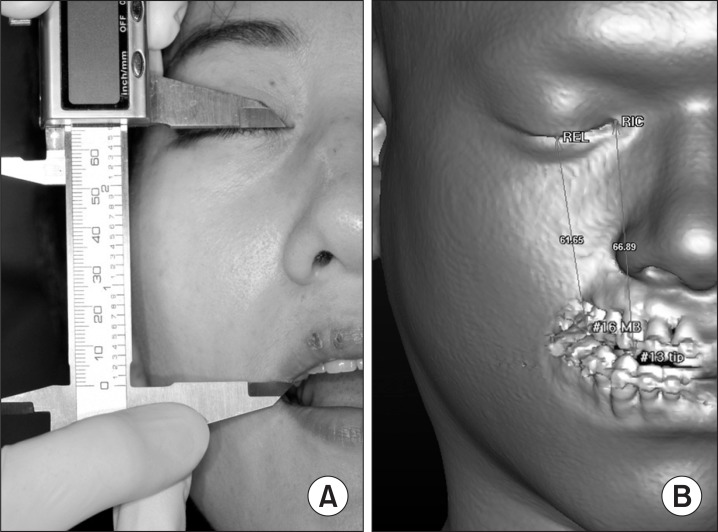
Fig. 2 A. Clinical measurement for the evaluation of occlusal cant. B. Virtual measurement on 3D CT.
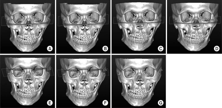
Fig. 3 Skeletal reference planes, line. A. FH plane R: Or.R - Or.L - Po.R; B. FH plane L: Or.R - Or.L - Po.L; C. F. Ovale plane L: Rt.F.Ovale - Lt.F.Ovale - Or.R; D. F. ovale plane L: Rt.F.Ovale - Lt.F.Ovale - Or.L; E. FZS plane R: Rt.FZS - Lt.FZS - Po.R; F. FZS plane R: Rt.FZS - Lt.FZS - Po.L; G. FZS line: Rt.FZS - Lt.FZS.
Reference
-
1. Proffit WR, White RP. Surgical-orthodontic treatment. St. Louis: Mosby-Year Book;1991.2. Lu KH. Harmonic analysis of the human face. Biometrics. 1965; 21:491–505. PMID: 14338681.
Article3. Peck S, Peck L, Kataja M. Skeletal asymmetry in esthetically pleasing faces. Angle Orthod. 1991; 61:43–48. PMID: 2012321.4. Ferrario VF, Sforza C, Poggio CE, Tartaglia G. Distance from symmetry: a three-dimensional evaluation of facial asymmetry. J Oral Maxillofac Surg. 1994; 52:1126–1132. PMID: 7965306.
Article5. Ferrario VF, Sforza C, Miani A, Tartaglia G. Craniofacial morphometry by photographic evaluations. Am J Orthod Dentofacial Orthop. 1993; 103:327–337. PMID: 8480698.
Article6. Padwa BL, Kaiser MO, Kaban LB. Occlusal cant in the frontal plane as a reflection of facial asymmetry. J Oral Maxillofac Surg. 1997; 55:811–816. PMID: 9251608.
Article7. McIntyre GT, Mossey PA. Posteroanterior cephalometric analysis of the parental craniofacial morphology in orofacial clefting. Cleft Palate Craniofac J. 2003; 40:416–425. PMID: 12846607.
Article8. Reyneke JP. Essentials of orthognathic surgery. 2nd ed. Hanover Park, IL: Quintessence Pub;2010.9. Polley JW, Figueroa AA, Liou EJ, Cohen M. Longitudinal analysis of mandibular asymmetry in hemifacial microsomia. Plast Reconstr Surg. 1997; 99:328–339. PMID: 9030137.
Article10. Cho BC, Shin DP, Park JW, Baik BS. Bimaxillary osteodistraction for the treatment of facial asymmetry in adults. Br J Plast Surg. 2001; 54:491–498. PMID: 11513510.
Article11. Rachmiel A, Manor R, Peled M, Laufer D. Intraoral distraction osteogenesis of the mandible in hemifacial microsomia. J Oral Maxillofac Surg. 2001; 59:728–733. PMID: 11429728.
Article12. Grayson B, Cutting C, Bookstein FL, Kim H, McCarthy JG. The three-dimensional cephalogram: theory, technique, and clinical application. Am J Orthod Dentofacial Orthop. 1988; 94:327–337. PMID: 3177285.13. Bergersen EO. Enlargement and distortion in cephalometric radiography: compensation tables for linear measurements. Angle Orthod. 1980; 50:230–244. PMID: 6931505.14. Ahlqvist J, Eliasson S, Welander U. The cephalometric projection. Part II. Principles of image distortion in cephalography. Dentomaxillofac Radiol. 1983; 12:101–108. PMID: 6584358.15. Habets LL, Bezuur JN, Naeiji M, Hansson TL. The Orthopantomogram, an aid in diagnosis of temporomandibular joint problems. II. The vertical symmetry. J Oral Rehabil. 1988; 15:465–471. PMID: 3244055.16. Türp JC, Vach W, Harbich K, Alt KW, Strub JR. Determining mandibular condyle and ramus height with the help of an Orthopantomogram--a valid method? J Oral Rehabil. 1996; 23:395–400. PMID: 8809694.17. Kusnoto B, Evans CA, BeGole EA, de Rijk W. Assessment of 3-dimensional computer-generated cephalometric measurements. Am J Orthod Dentofacial Orthop. 1999; 116:390–399. PMID: 10511666.
Article18. Cavalcanti MG, Ruprecht A, Bonomie JM, Vannier MW. The validation of 3D spiral CT-based measurements of simulated maxillofacial neoplasms. Oral Surg Oral Med Oral Pathol Oral Radiol Endod. 2000; 89:753–758. PMID: 10846133.19. Fuhrmann RA, Schnappauf A, Diedrich PR. Three-dimensional imaging of craniomaxillofacial structures with a standard personal computer. Dentomaxillofac Radiol. 1995; 24:260–263. PMID: 9161172.
Article20. Matteson SR, Bechtold W, Phillips C, Staab EV. A method for three-dimensional image reformation for quantitative cephalometric analysis. J Oral Maxillofac Surg. 1989; 47:1053–1061. PMID: 2795298.
Article21. Hildebolt CF, Vannier MW, Knapp RH. Validation study of skull three-dimensional computerized tomography measurements. Am J Phys Anthropol. 1990; 82:283–294. PMID: 2375381.
Article22. Hassan B, Nijkamp P, Verheij H, Tairie J, Vink C, van der Stelt P, et al. Precision of identifying cephalometric landmarks with cone beam computed tomography in vivo. Eur J Orthod. 2013; 35:38–44. PMID: 21447781.
Article23. Katsumata A, Fujishita M, Maeda M, Ariji Y, Ariji E, Langlais RP. 3D-CT evaluation of facial asymmetry. Oral Surg Oral Med Oral Pathol Oral Radiol Endod. 2005; 99:212–220. PMID: 15660095.
Article24. Hwang HS, Hwang CH, Lee KH, Kang BC. Maxillofacial 3-dimensional image analysis for the diagnosis of facial asymmetry. Am J Orthod Dentofacial Orthop. 2006; 130:779–785. PMID: 17169741.
Article25. Bell WH. Modern practice in orthognathic and reconstructive surgery. Philadelphia: Saunders;1992.
- Full Text Links
-
- Actions
-
Cited
- CITED
-
- Close
- Share
-
- Similar articles
-
- A study on horizontal reference planes in lateral cephalogram in Korean children
- A study on horizontal reference planes in lateral cephalogram in Korean adults
- The relationship among reference lines used for taking the extraoral radiography
- Correlation between Cephalometric Reference Planes for Clinical Application to Articulators
- Use of the orbito-occipital line as an alternative to the Frankfort line
