Diffusion-Weighted Magnetic Resonance Imaging Findings in a Patient with Trigeminal Ganglioneuroma
- Affiliations
-
- 1Department of Radiology, Chonnam National University Medical School, Chonnam National University Hospital, Gwangju 501-757, Korea. radyoon@jnu.ac.kr
- KMID: 1430054
- DOI: http://doi.org/10.3348/kjr.2013.14.1.118
Abstract
- A case of intracranial ganglioneuroma arising from the trigeminal nerve in the pontine and cerebellopontine angle cistern, in a 44-year-old female, is presented with an emphasis on diffusion-weighted imaging findings. We will discuss on how the tumor in the very unusual location should be differentiated particularly focused on diffusion-weighted imaging findings.
MeSH Terms
Figure
-
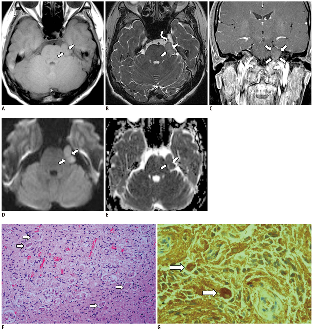
Fig. 1 Trigeminal ganglioneuroma in 44-year-old female. A. Axial T1-weighted image shows well-defined mass (arrows) in left prepontine and cerebellopontine angle cistern. This has same signal intensity as adjacent brain parenchyma. B. Thin section heavily T2-weighted image shows mass (arrows) with abutting and displacement of left trigeminal nerve (curved arrow). C. Coronal postcontrast T1-weighted image shows minimal contrast enhancement of tumor (arrows). D. Axial echo-planar diffusion-weighed imaging with b = 1000 s/mm2 shows tumor with hyperintense lesion relative to adjacent brain parenchyma. E. Apparent diffusion coefficient map shows mild hypointensity of tumor (arrows). F. Photomicrograph shows that tumor consists of scattered mature ganglion cells (arrows) and spindle-shaped cells with scanty myxoid changes, H&E stain, × 200. G. Photomicrograph shows relatively strong S-100 expression of ganglion cells (arrows), × 400.
Cited by 1 articles
-
Use of Magnetic Resonance Neurography for Evaluating the Distribution and Patterns of Chronic Inflammatory Demyelinating Polyneuropathy
Xiaoyun Su, Xiangquan Kong, Zuneng Lu, Min Zhou, Jing Wang, Xiaoming Liu, Xiangchuang Kong, Huiting Zhang, Chuansheng Zheng
Korean J Radiol. 2020;21(4):483-493. doi: 10.3348/kjr.2019.0739.
Reference
-
1. Abe T, Asano T, Manabe T, Matsuura H, Furuta T, Taguchi K. Trigeminal ganglioneuroma. Brain Tumor Pathol. 1999. 16:49–53.2. Arseni C, Horvath L, Carp N, Ciurea V. Intracranial ganglioneuromas in children. Acta Neurochir (Wien). 1975. 32:279–286.3. Lonergan GJ, Schwab CM, Suarez ES, Carlson CL. Neuroblastoma, ganglioneuroblastoma, and ganglioneuroma: radiologic-pathologic correlation. Radiographics. 2002. 22:911–934.4. Cai J, Zeng Y, Zheng H, Qin Y, T K, Zhao J. Retroperitoneal ganglioneuroma in children: CT and MRI features with histologic correlation. Eur J Radiol. 2010. 75:315–320.5. Zhang Y, Nishimura H, Kato S, Fujimoto K, Ohkuma K, Kojima K, et al. MRI of ganglioneuroma: histologic correlation study. J Comput Assist Tomogr. 2001. 25:617–623.6. Gahr N, Darge K, Hahn G, Kreher BW, von Buiren M, Uhl M. Diffusion-weighted MRI for differentiation of neuroblastoma and ganglioneuroblastoma/ganglioneuroma. Eur J Radiol. 2011. 79:443–446.7. Qing Y, Bin X, Jian W, Li G, Linhui W, Bing L, et al. Adrenal ganglioneuromas: a 10-year experience in a Chinese population. Surgery. 2010. 147:854–860.8. Mermuys K, Wilms G, Demaerel P. Epidermoid cyst of the fourth ventricle: diffusion-weighted and flair MR imaging findings. JBR-BTR. 2008. 91:58–60.9. Castro DE, Raghuram K, Phillips CD. Benign triton tumor of the trigeminal nerve. AJNR Am J Neuroradiol. 2005. 26:967–969.10. Athale S, Hallet KK, Jinkins JR. Ganglioglioma of the trigeminal nerve: MRI. Neuroradiology. 1999. 41:576–578.
- Full Text Links
-
- Actions
-
Cited
- CITED
-
- Close
- Share
-
- Similar articles
-
- RE: Diffusion-Weighted Imaging of Prostate Cancer: How Can We Use It Accurately?
- Squamous Cell Carcinoma of the Pancreas: A Case Report
- The Value of PROPELLER Diffusion-Weighted Image in the Detection of Cholesteatoma
- Diffusion-Weighted Magnetic Resonance Imaging of Spine
- Clear Cell Sarcoma of the Wrist: MRI Findings with Diffusion-Weighted Image and Histopathologic Correlation
