New Landmark for the Endoscopic Endonasal Transsphenoidal Approach of Pituitary Surgery
- Affiliations
-
- 1Department of Otorhinolaryngology-Head and Neck Surgery, The Catholic University of Korea College of Medicine, Seoul, Korea. entcho@catholic.ac.kr
- KMID: 1426207
- DOI: http://doi.org/10.3340/jkns.2013.53.4.218
Abstract
OBJECTIVE
To clarify the anatomical correlations of the sphenoid sinus with surrounding structures in the normal Korean population, and to identify surgical landmarks for safe sellar floor dissection in the anterior skull base by endoscopy and microscopy.
METHODS
We reviewed the 196 brain magnetic resonance imaging findings showing a normal appearance, and measured the distances between anatomical landmarks.
RESULTS
The mean distances from the base of the columella to the anterior wall of the sphenoid sinus and the sellar floor were 69.71+/-4.25 mm and 86.26+/-4.57 mm, respectively in the over 15 age group, and showed the smallest degree of variation among the measurements. The mean angles between the floor of the nasal cavity and the straight line connecting the base of the columella and the sellar floor were 29.45+/-3.25degrees and 24.75+/-4.00degrees in the over 15 and under 15 age groups, respectively. The mean values of both distances and angles increased with age until 15 years after which no further increases were evident. There were no significant differences in the measurements between males and females or among subjects with different degrees of pneumatization in the over 15 age group.
CONCLUSION
The distances from the base of the columella to the sellar floor and the anterior wall of the sphenoid sinus, which were consistent among individuals, could be used as a surgical indicator to investigate the sellar floor in endoscopic or microscopic transsphenoidal approaches.
MeSH Terms
Figure
-
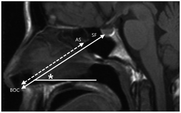
Fig. 1 Measurement of the distances between the base of the columella (BOC) and the anterior wall of the sphenoid sinus (AS) and between the BOC and the sellar floor (SF) on T1 weighted fast spin-echo sagittal images of the brain. We measured the line drawn between the BOC and the SF (double arrowed line) and determined the shortest distance between the BOC and the AS (dashed line) on that line. In addition, the angle (asterisk) between the nasal cavity floor and the line connecting the BOC and the SF was measured.
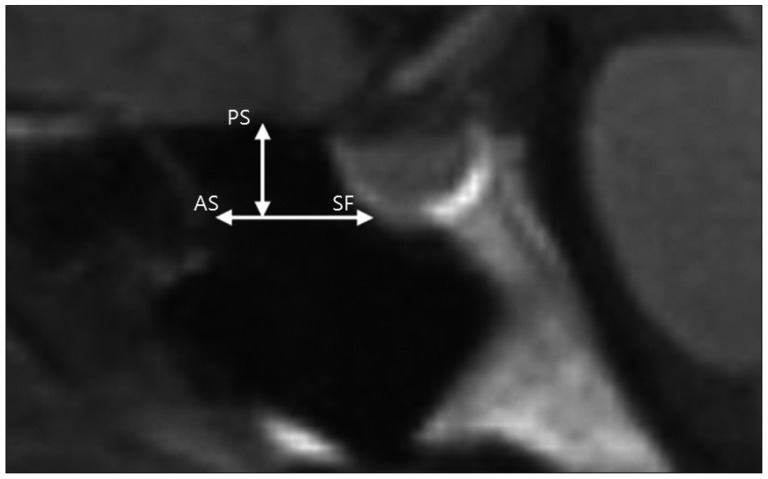
Fig. 2 Measurement of the anteroposterior distances between the anterior wall of the sphenoid sinus (AS) and the sellar floor (SF) and the craniocaudal distances between the planum sphenoidale (PS) and the SF on T1 weighted fast spin-echo sagittal images of the brain. We chose one point on the SF at which the perpendicular bony wall from the tuberculum sella turns horizontally, and measured the distance between the AS and the SF (horizontal line) and determined the shortest distance between the horizontal line and the PS (vertical line).
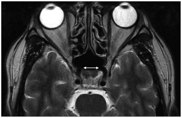
Fig. 3 The shortest distances between both internal carotid arteries and the anterior-most portions of the cavernous segment (C4) were measured on T2 weighted fast spin-echo axial images of the brain (double arrow).
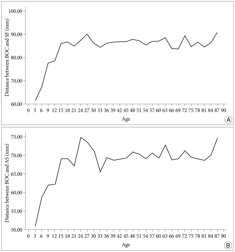
Fig. 4 Mean values of the distance in patients grouped according to age at intervals of 3 years. A : Changes in distance between the base of the columella (BOC) and the sellar floor (SF) with age. B : Change in distance between the BOC and the anterior wall of the sphenoid sinus (AS) in relation to age. Mean values of the distance increased until age 15 and then showed a plateau.
Cited by 1 articles
-
Feasibility of Endoscopic Endonasal Approach for Recurrent Pituitary Adenomas after Microscopic Trans-Sphenoidal Approach
Joo Min Hwang, Yong Hwy Kim, Jin Wook Kim, Dong Gyu Kim, Hee-Won Jung, Young Seob Chung
J Korean Neurosurg Soc. 2013;54(4):317-322. doi: 10.3340/jkns.2013.54.4.317.
Reference
-
1. Barghouth G, Prior JO, Lepori D, Duvoisin B, Schnyder P, Gudinchet F. Paranasal sinuses in children : size evaluation of maxillary, sphenoid, and frontal sinuses by magnetic resonance imaging and proposal of volume index percentile curves. Eur Radiol. 2002; 12:1451–1458. PMID: 12042953.
Article2. Campero A, Socolovsky M, Torino R, Martins C, Yasuda A, Rhoton AL Jr. Anatomical landmarks for positioning the head in preparation for the transsphenoidal approach : the spheno-sellar point. Br J Neurosurg. 2009; 23:282–286. PMID: 19533460.
Article3. Cavallo LM, Messina A, Cappabianca P, Esposito F, de Divitiis E, Gardner P, et al. Endoscopic endonasal surgery of the midline skull base : anatomical study and clinical considerations. Neurosurg Focus. 2005; 19:E2. PMID: 16078816.4. Cushing H. III. Partial Hypophysectomy for Acromegaly : with Remarks on the Function of the Hypophysis. Ann Surg. 1909; 50:1002–1017. PMID: 17862444.
Article5. Enatsu K, Takasaki K, Kase K, Jinnouchi S, Kumagami H, Nakamura T, et al. Surgical anatomy of the sphenoid sinus on the CT using multiplanar reconstruction technique. Otolaryngol Head Neck Surg. 2008; 138:182–186. PMID: 18241713.
Article6. Gulsen S, Dinc AH, Unal M, Cantürk N, Altinors N. Characterization of the anatomic location of the pituitary stalk and its relationship to the dorsum sellae, tuberculum sellae and chiasmatic cistern. J Korean Neurosurg Soc. 2010; 47:169–173. PMID: 20379467.
Article7. Hamid O, El Fiky L, Hassan O, Kotb A, El Fiky S. Anatomic Variations of the Sphenoid Sinus and Their Impact on Trans-sphenoid Pituitary Surgery. Skull Base. 2008; 18:9–15. PMID: 18592020.
Article8. Hatipoglu HG, Cetin MA, Selvi A, Yuksel E. Role of magnetic resonance imaging in evaluating sphenoid sinus and internal carotid artery. J Laryngol Otol. 2009; 123:1331–1337. PMID: 19775490.
Article9. Jane JA Jr, Han J, Prevedello DM, Jagannathan J, Dumont AS, Laws ER Jr. Perspectives on endoscopic transsphenoidal surgery. Neurosurg Focus. 2005; 19:E2. PMID: 16398479.
Article10. Jang YJ, Kim SC. Pneumatization of the sphenoid sinus in children evaluated by magnetic resonance imaging. Am J Rhinol. 2000; 14:181–185. PMID: 10887625.
Article11. Ju KS, Bae HG, Park HK, Chang JC, Choi SK, Sim KB. Morphometric study of the Korean adult pituitary glands and the diaphragma sellae. J Korean Neurosurg Soc. 2010; 47:42–47. PMID: 20157377.
Article12. Kim HU, Kim SS, Kang SS, Chung IH, Lee JG, Yoon JH. Surgical anatomy of the natural ostium of the sphenoid sinus. Laryngoscope. 2001; 111:1599–1602. PMID: 11568612.
Article
- Full Text Links
-
- Actions
-
Cited
- CITED
-
- Close
- Share
-
- Similar articles
-
- Deliberate Two-Staged Endoscopic Endonasal Transsphenoidal Surgery in Large Pituitary Adenomas
- Endoscopic Endonasal Surgery for Pituitary Adenoma
- Clinical Analysis of Endoscopic Transnasal Transsphenoidal Hypophysectomy of Pituitary Tumor
- Endoscopic Endonasal Transsphenoidal Pituitary Tumor Surgery: An Early Experience
- Endocrine Outcome of Endoscopic Endonasal Transsphenoidal Surgery in Functioning Pituitary Adenomas
