J Korean Orthop Assoc.
2013 Apr;48(2):89-95. 10.4055/jkoa.2013.48.2.89.
Supramalleolar Tibial Osteotomy for Medial Compartment Ankle Osteoarthritis
- Affiliations
-
- 1Department of Orthopedic Surgery, Konkuk University School of Medicine, Seoul, Korea. jungfoot@hanmail.net
- KMID: 1424092
- DOI: http://doi.org/10.4055/jkoa.2013.48.2.89
Abstract
- PURPOSE
The aim of this study is to evaluate the clinical and radiologic outcomes of supramalleolar tibial osteotomy for medial compartment ankle osteoarthritis (OA) and to verify the efficacy of the supramalleolar osteotomy.
MATERIALS AND METHODS
This study is based on 9 ankles of the medial compartment ankle OA treated with supramalleolar tibial osteotomy from August 2007 to June 2011 with at least 1 year follow-up. As for the functional evaluation, visual analogue scale (VAS) pain scores and American Orthopaedic Foot and Ankle Society (AOFAS) ankle-hindfoot scores were evaluated. On radiographs, tibial anterior surface (TAS) angles, tibial lateral surface angles were measured. The severity of ankle OA was classified by the Takaura staging system.
RESULTS
The mean VAS pain scores improved to 0.6 and AOFAS scores improved to 89.3. Radiographically, TAS angle increased to 93.5degrees postoperatively. Seven ankles showed improvement of the ankle arthritis grading from IIIa to II according to Takakura's staging.
CONCLUSION
Supramalleolar tibial osteotomy for patients with medial compartment varus ankle OA showed satisfactory clinical and radiological outcome. We confirmed that the procedure is recommendable for medial compartment varus ankle OA especially for Takakura stage IIIa.
Figure
-
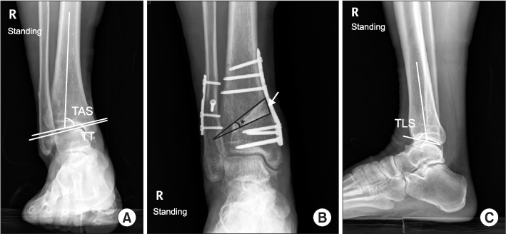
Figure 1 (A) Anteroposterior view demonstrates measurement of tibial anterior surface angle (TAS) and talar tilt angle (TT). (B) Measurement of wedge angle (astrix) and wedge height (arrow). (C) Lateral view demonstrates measurement of tibial lateral surface angle (TLS).
Figure 2 A 55-year-old woman with medial compartment ankle osteoarthritis. (A) Preoperative standing radiograph (B) treated by supramalleolar tibial and fibula osteotomies, which resulted in an excellent functional outcome.
Cited by 1 articles
-
Low Tibial Osteotomy
Jun Young Choi, Jin Soo Suh
J Korean Foot Ankle Soc. 2014;18(4):141-146. doi: 10.14193/jkfas.2014.18.4.141.
Reference
-
1. Valderrabano V, Horisberger M, Russell I, Dougall H, Hintermann B. Etiology of ankle osteoarthritis. Clin Orthop Relat Res. 2009. 467:1800–1806.
Article2. Thomas RH, Daniels TR. Ankle arthritis. J Bone Joint Surg Am. 2003. 85:923–936.
Article3. Lee HS, Wapner KL, Park SS, Kim JS, Lee DH, Sohn DW. Ligament reconstruction and calcaneal osteotomy for osteoarthritis of the ankle. Foot Ankle Int. 2009. 30:475–480.
Article4. Takakura Y, Tanaka Y, Kumai T, Tamai S. Low tibial osteotomy for osteoarthritis of the ankle. Results of a new operation in 18 patients. J Bone Joint Surg Br. 1995. 77:50–54.
Article5. Stamatis ED, Cooper PS, Myerson MS. Supramalleolar osteotomy for the treatment of distal tibial angular deformities and arthritis of the ankle joint. Foot Ankle Int. 2003. 24:754–764.
Article6. Pagenstert GI, Hintermann B, Barg A, Leumann A, Valderrabano V. Realignment surgery as alternative treatment of varus and valgus ankle osteoarthritis. Clin Orthop Relat Res. 2007. 462:156–168.
Article7. Tanaka Y, Takakura Y, Hayashi K, Taniguchi A, Kumai T, Sugimoto K. Low tibial osteotomy for varus-type osteoarthritis of the ankle. J Bone Joint Surg Br. 2006. 88:909–913.
Article8. Warnock KM, Johnson BD, Wright JB, Ambrose CG, Clanton TO, McGarvey WC. Calculation of the opening wedge for a low tibial osteotomy. Foot Ankle Int. 2004. 25:778–782.
Article9. Takakura Y, Takaoka T, Tanaka Y, Yajima H, Tamai S. Results of opening-wedge osteotomy for the treatment of a post-traumatic varus deformity of the ankle. J Bone Joint Surg Am. 1998. 80:213–218.
Article10. Cheng YM, Huang PJ, Hong SH, et al. Low tibial osteotomy for moderate ankle arthritis. Arch Orthop Trauma Surg. 2001. 121:355–358.
Article11. Knupp M, Stufkens SA, van Bergen CJ, et al. Effect of supramalleolar varus and valgus deformities on the tibiotalar joint: a cadaveric study. Foot Ankle Int. 2011. 32:609–615.
Article12. Tarr RR, Resnick CT, Wagner KS, Sarmiento A. Changes in tibiotalar joint contact areas following experimentally induced tibial angular deformities. Clin Orthop Relat Res. 1985. 199:72–80.
Article13. Lee WC, Moon JS, Lee K, Byun WJ, Lee SH. Indications for supramalleolar osteotomy in patients with ankle osteoarthritis and varus deformity. J Bone Joint Surg Am. 2011. 93:1243–1248.
Article14. Moon JS, Shim JC, Suh JS, Lee WC. Radiographic predictability of cartilage damage in medial ankle osteoarthritis. Clin Orthop Relat Res. 2010. 468:2188–2197.
Article15. Lee WC, Moon JS, Lee HS, Lee K. Alignment of ankle and hindfoot in early stage ankle osteoarthritis. Foot Ankle Int. 2011. 32:693–699.
Article
- Full Text Links
-
- Actions
-
Cited
- CITED
-
- Close
- Share
-
- Similar articles
-
- Supramalleolar Distal Tibiofibular Osteotomy for Medial Ankle Osteoarthritis: Current Concepts
- Supramalleolar Osteotomy for Moderate Degenerative Ankle Osteoarthritis
- Supramalleolar Osteotomy in Patients with Varus Ankle Osteoarthritis
- Low Tibial Osteotomy
- Supramalleolar Osteotomy Combined with Redo Arthroscopy for a Patient with Persistent Pain after Primary Arthroscopic Microfracture for Medial Osteochondral Lesion of the Talus: A Case Report
