Supramalleolar Osteotomy Combined with Redo Arthroscopy for a Patient with Persistent Pain after Primary Arthroscopic Microfracture for Medial Osteochondral Lesion of the Talus: A Case Report
- Affiliations
-
- 1Department of Orthopedic Surgery, Ilsan Paik Hospital, Inje University College of Medicine, Goyang, Korea
- KMID: 2543004
- DOI: http://doi.org/10.14193/jkfas.2023.27.2.71
Abstract
- A medial opening wedge supramalleolar osteotomy (SMO) introduced by Takakura et al. is a useful realignment procedure for patients with ankle joint arthritis and varus malalignment by shifting the weight-bearing axis laterally and redistributing the loads on the ankle joint. When pain persists after arthroscopic microfracture in patients with medial osteochondral lesion of the talus (OLT), redo arthroscopy, osteochondral autograft transplantation, autologous chondrocyte implantation, or matrix-induced chondrogenesis might be indicated. On the other hand, there is insufficient scientific evidence for realignment surgery through SMO, while the effect of realignment surgery has been studied consecutively for osteochondral lesions of the knee. Therefore, this paper reports a patient with medial OLT who underwent redo arthroscopy combined with SMO for persistent pain after primary arthroscopic microfracture.
Figure
-
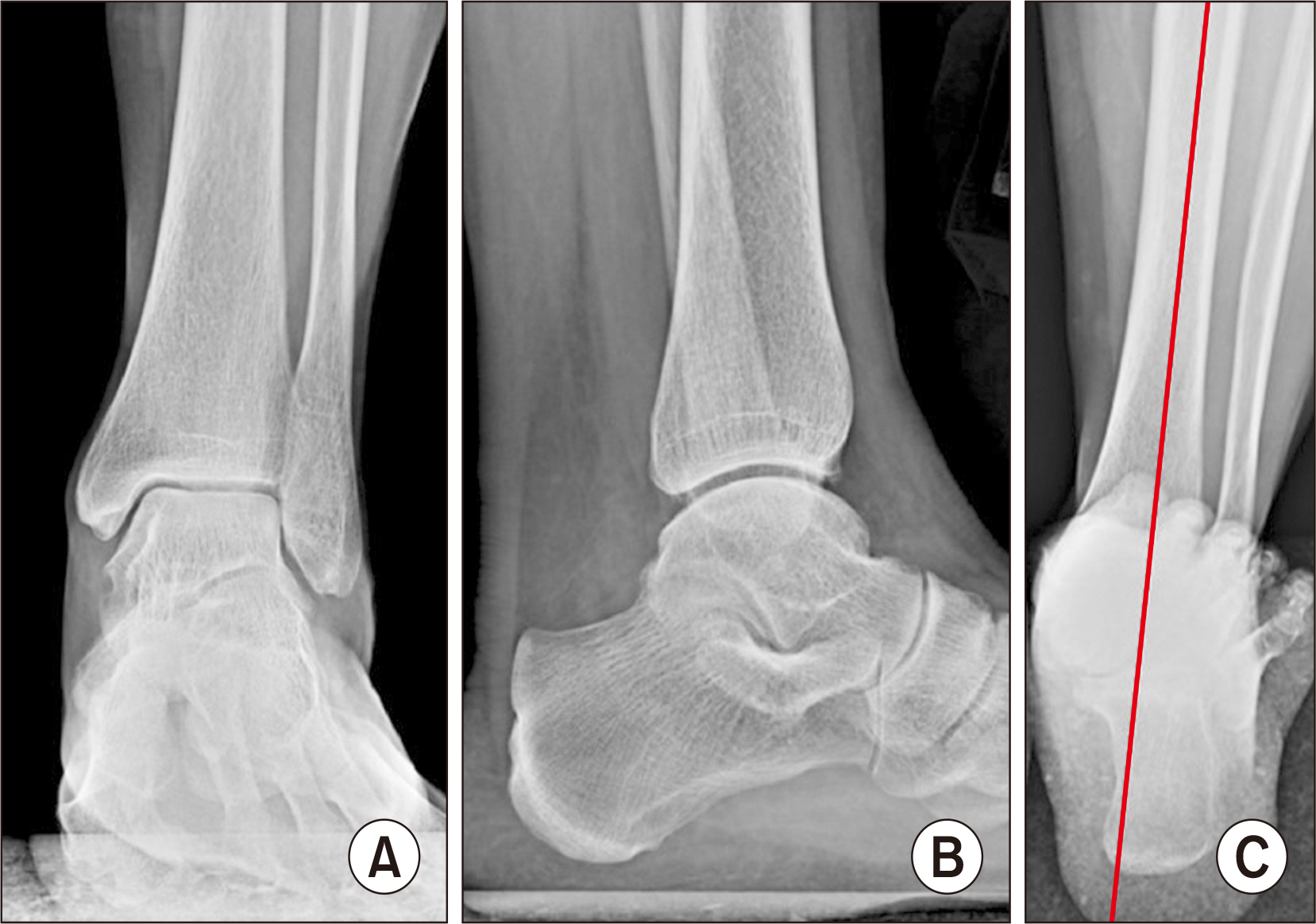
Figure 1 On standing anteroposterior (A) and lateral (B) radiographs, no specific abnormal findings were found while the hindfoot valgus alignment was noted on hindfoot alignment view (C).
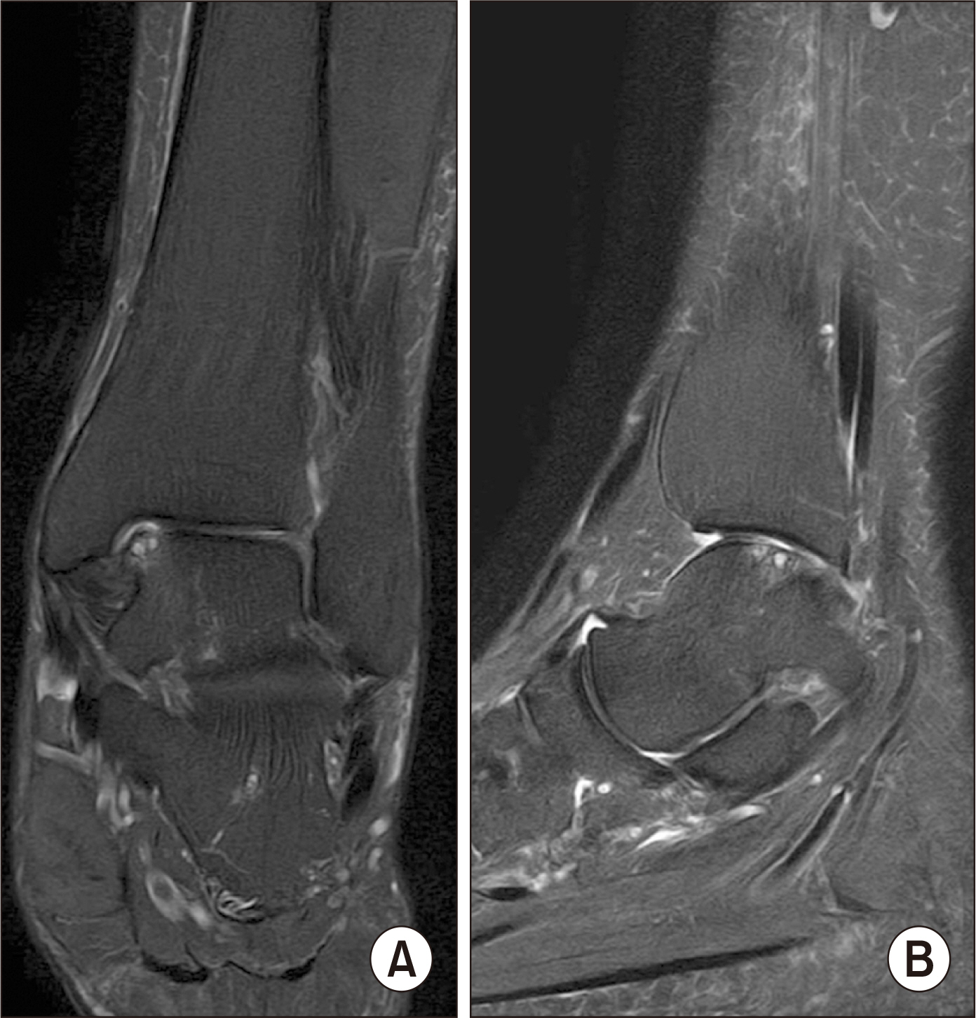
Figure 2 On T2-weighted coronal (A) and sagittal (B) magnetic resonance imaging, subchondral cyst formations were found with a suspicious cartilage defect on medial talar dome.
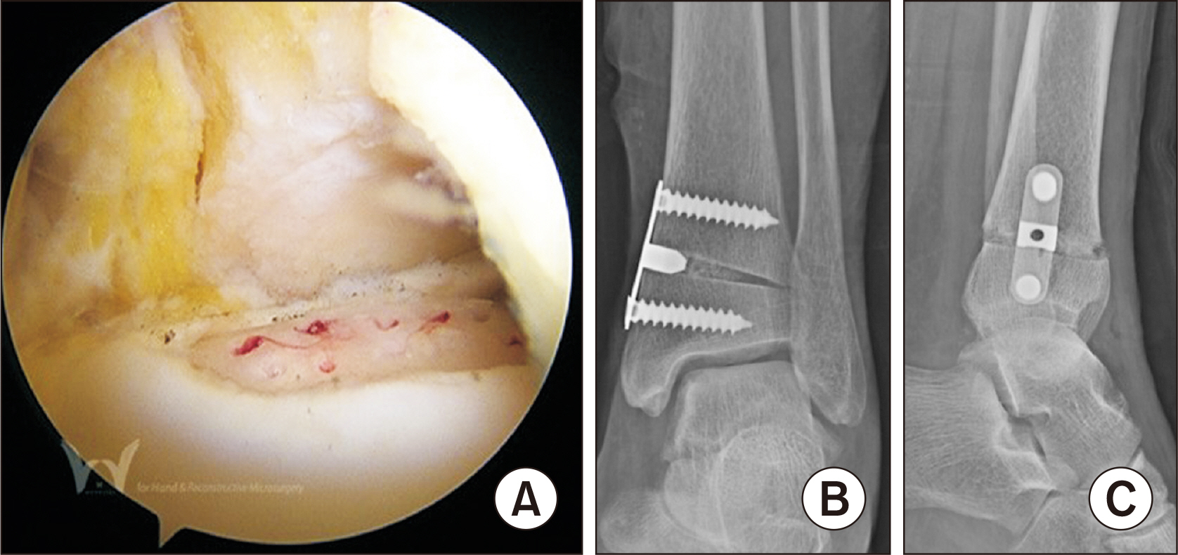
Figure 3 Redo arthroscopic microfracture was performed (A) followed by medial opening wedge supramalleolar osteotomy with 6 mm wedge (B: anteroposterior view, C: lateral view).
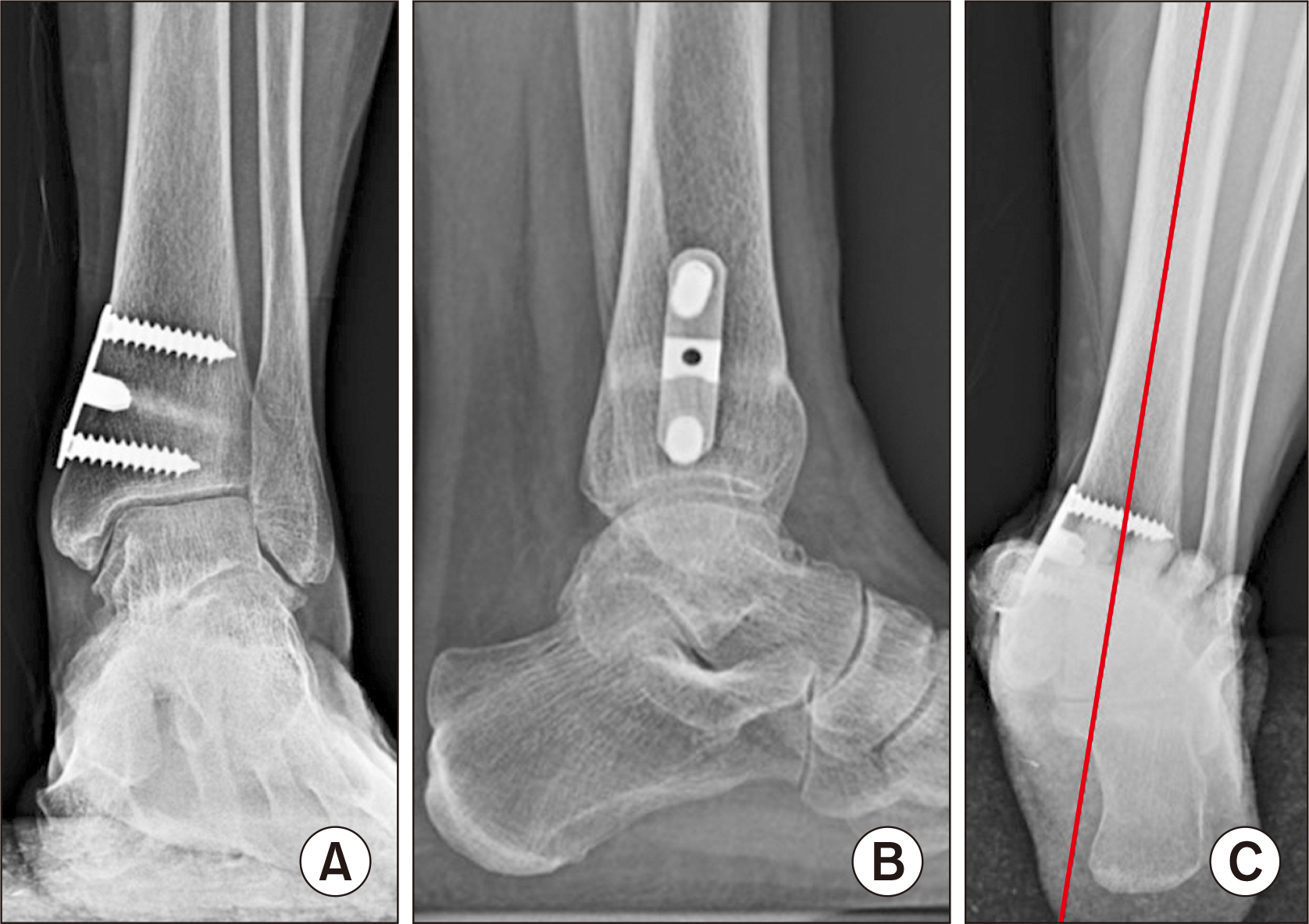
Figure 4 At postoperative 41 months, patient’s symptom was subsided with a stable maintenance of osteotomy site without a further deterioration of prior osteochondral lesion of talus (A: anteroposterior view, B: lateral view, C: hindfoot alignment view).
Reference
-
1. Choi WJ, Park KH, Lee M, Chung K, Lee JW. 2015; Redomicrofracture as a treatment for osteochondral lesion of talus after the failure of arthroscopic microfracture. J Korean Foot Ankle Soc. 19:43–6. doi: 10.14193/jkfas.2015.19.2.43. DOI: 10.14193/jkfas.2015.19.2.43.
Article2. Yoon HS, Park YJ, Lee M, Choi WJ, Lee JW. 2014; Osteochondral autologous transplantation is superior to repeat arthroscopy for the treatment of osteochondral lesions of the talus after failed primary arthroscopic treatment. Am J Sports Med. 42:1896–903. doi: 10.1177/0363546514535186. DOI: 10.1177/0363546514535186. PMID: 24907287.
Article3. Kim JS. 2015; Autologous chondrocyte implantation as a secondary procedure after failed microfracture for osteochondral lesion of talus. J Korean Foot Ankle Soc. 19:7–10. doi: 10.14193/jkfas.2015.19.1.7. DOI: 10.14193/jkfas.2015.19.1.7.
Article4. Kim BS, Na Y, Kwon WH. 2020; Operative treatment for osteochondral lesions of the talus: bone marrow aspirate concentrate and matrix-induced chondrogenesis. J Korean Foot Ankle Soc. 24:61–8. doi: 10.14193/jkfas.2020.24.2.61. DOI: 10.14193/jkfas.2020.24.2.61.
Article5. Mina C, Garrett WE Jr, Pietrobon R, Glisson R, Higgins L. 2008; High tibial osteotomy for unloading osteochondral defects in the medial compartment of the knee. Am J Sports Med. 36:949–55. doi: 10.1177/0363546508315471. DOI: 10.1177/0363546508315471. PMID: 18413679.
Article6. Agarwalla A, Christian DR, Liu JN, Garcia GH, Redondo ML, Gowd AK, et al. 2020; Return to work following high tibial osteotomy with concomitant osteochondral allograft transplantation. Arthroscopy. 36:808–15. doi: 10.1016/j.arthro.2019.08.046. DOI: 10.1016/j.arthro.2019.08.046. PMID: 31870751.
Article7. Savva N, Jabur M, Davies M, Saxby T. 2007; Osteochondral lesions of the talus: results of repeat arthroscopic debridement. Foot Ankle Int. 28:669–73. doi: 10.3113/FAI.2007.0669. DOI: 10.3113/FAI.2007.0669. PMID: 17592696.
Article8. Arshad Z, Aslam A, Iqbal AM, Bhatia M. 2022; Should arthroscopic bone marrow stimulation be used in the management of secondary osteochondral lesions of the talus? A systematic review. Clin Orthop Relat Res. 480:1112–25. doi: 10.1097/CORR.0000000000002134. DOI: 10.1097/CORR.0000000000002134. PMID: 35130190. PMCID: PMC9263474.
Article9. Li X, Zhu Y, Xu Y, Wang B, Liu J, Xu X. 2017; Osteochondral autograft transplantation with biplanar distal tibial osteotomy for patients with concomitant large osteochondral lesion of the talus and varus ankle malalignment. BMC Musculoskelet Disord. 18:23. doi: 10.1186/s12891-016-1367-2. DOI: 10.1186/s12891-016-1367-2. PMID: 28103870. PMCID: PMC5244526.
Article10. Kim YM, Kim KI, Han B. 2020; If the lower extremity alignment is corrected, will osteochondral lesions of the talus improve? J Korean Foot Ankle Soc. 24:42–7. doi: 10.14193/jkfas.2020.24.2.42. DOI: 10.14193/jkfas.2020.24.2.42.
Article
- Full Text Links
-
- Actions
-
Cited
- CITED
-
- Close
- Share
-
- Similar articles
-
- Redomicrofracture as a Treatment for Osteochondral Lesion of Talus after the Failure of Arthroscopic Microfracture
- Autologous Osteochondral Transplantation as a Secondary Procedure after Failed Microfracture for Osteochondral Lesion of Talus
- Autologous Chondrocyte Implantation as a Secondary Procedure after Failed Microfracture for Osteochondral Lesion of Talus
- If the Patient Complains Persistent Pain after the Operation, What Should We Do?
- Second-look Arthroscopy after Surgical Treatment for Osteochondral Lesion of Talus: Comparison of Mosaicplasty with Microfracture
