Endoscope-Assisted Microsurgical Removal of an Epidermoid Tumor within the Cavernous Sinus
- Affiliations
-
- 1Department of Neurosurgery, Eulji University Hospital, Daejeon, Korea. nsksm@eulji.ac.kr
- 2Brain Tumor and Neuro-oncology Center, Department of Neurosurgery, Cleveland Clinic, Cleveland, OH, USA.
- KMID: 1414267
- DOI: http://doi.org/10.3349/ymj.2012.53.6.1216
Abstract
- Epidermoid tumor of the cavernous sinus is rare. The aim of this case report is to discuss the role of neuroendoscopes in the removal of such lesions. A 21-year-old man presented with 6-year history of progressive headache, diplopia, and visual disturbance. Work-up revealed an epidermoid tumor located in the right cavernous sinus. An extradural transcavernous approach was utilized via a traditional frontotemporal craniotomy with endoscopic assistance. The postoperative course was uneventful with immediate improvement of the patient's headache. Postoperative magnetic resonance imaging demonstrated complete removal of the tumor. There were no signs of recurrence during a 2-year follow-up period. The endoscope is a useful tool for removing epidermoid tumors from the cavernous sinus and enhances visualization of areas that would otherwise be difficult to visualize with microscopes alone. Endoscopes also help minimize the retraction of neurovascular structures.
MeSH Terms
Figure
-

Fig. 1 (A) The extradural transcavernous approach, peeling the outer dural layer away from the lateral wall of the cavernous sinus. (B and C) Exposure of the yellowish-white tumor following a small incision in the membranous layer of the lateral wall of the cavernous sinus. (D) The advancement of a 4 mm rigid neuroendoscope inside the cavernous sinus through the small incision. (E) 0°-angled endoscopic visualization of a glistening white cauliflower-like mass adherent to the inner layer of the wall. (F) Following removal of the tumor, the cavernous segment of the internal carotid artery became visible, under a 30°-angled view. SOF, superior orbital fissure; FD, frontal dura; TD, temporal dura; IL, inner membranous layer of lateral cavernous sinus wall; T, epidermoid tumor; E, shaft of neuroendoscope; S, shaft of suction; P, anterior petroclinoidal fold; C, thinned trochlear nerve (dotted lines); I, cavernous segment of the internal carotid artery (dashed lines).

Fig. 2 (A) Preoperative magnetic resonance image demonstrates space-occupying slight hyper signal intensity within the right cavernous sinus and more bright lesions on diffusion-weighted images. (B) Magnetic resonance imaging performed on the first postoperative day showing complete removal of the tumor and signal change.
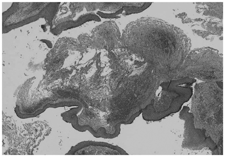
Fig. 3 Histopathologic examination revealed a cystic tumor lined with simple stratified keratinizing squamous epithelium.
Reference
-
1. Gharabaghi A, Koerbel A, Samii A, Safavi-Abbasi S, Tatagiba M, Samii M. Epidermoid cysts of the cavernous sinus. Surg Neurol. 2005. 64:428–433.
Article2. Tatagiba M, Iaconetta G, Samii M. Epidermoid cyst of the cavernous sinus: clinical features, pathogenesis and treatment. Br J Neurosurg. 2000. 14:571–575.
Article3. Bonde V, Goel A. Interdural cavernous sinus epidermoid cyst. J Clin Neurosci. 2008. 15:212–214.
Article4. Iaconetta G, Carvalho GA, Vorkapic P, Samii M. Intracerebral epidermoid tumor: a case report and review of the literature. Surg Neurol. 2001. 55:218–222.
Article5. Ikezaki K, Toda K, Abe M, Tabuchi K. Intracavernous epidermoid tumor presenting with abducens nerve paresis--case report. Neurol Med Chir (Tokyo). 1992. 32:360–364.
Article6. Cobbs CS, Pitts LH, Wilson CB. Epidermoid and dermoid cysts of the posterior fossa. Clin Neurosurg. 1997. 44:511–528.7. Kaido T, Okazaki A, Kurokawa S, Tsukamoto M. Pathogenesis of intraparenchymal epidermoid cyst in the brain: a case report and review of the literature. Surg Neurol. 2003. 59:211–216.8. Sekhar LN, Sen CN, Jho HD, Janecka IP. Surgical treatment of intracavernous neoplasms: a four-year experience. Neurosurgery. 1989. 24:18–30.
Article9. Fries G, Perneczky A. Endoscope-assisted brain surgery: part 2--analysis of 380 procedures. Neurosurgery. 1998. 42:226–231.
Article
- Full Text Links
-
- Actions
-
Cited
- CITED
-
- Close
- Share
-
- Similar articles
-
- Direct Microsurgical Repair of Traumatic Carotid-Cavernous Fistula
- Imaging Findings of a Nonenhancing Intradural Paramedian Chordoma Mimicking an Epidermoid Cyst
- Surgical Extent of Transsphenoidal Approach: A Microsurgical Anatomy
- The Outcome of Surgery Treatment of Cranial Base Meningiomas
- Pure Intracavernous Sinus Epidermoid Cyst: Diffusion-Weighted (DW) and Constructive Interference in Steady State (CISS) Images
