Pure Intracavernous Sinus Epidermoid Cyst: Diffusion-Weighted (DW) and Constructive Interference in Steady State (CISS) Images
- Affiliations
-
- 1Department of Radiology, Eulji University Hospital, Daejeon, Korea. midosyu@eulji.ac.kr
- 2Department of Neurosurgery, Eulji University Hospital, Daejeon, Korea.
- KMID: 2208879
- DOI: http://doi.org/10.3348/jksr.2010.63.1.9
Abstract
- An epidermoid cyst located in the true intracavernous sinus is extremely rare. A 22-year-old man presented with right ophthalmoplegia that had developed 3 years previously. The computed tomography (CT) and magnetic resonance (MR) images demonstrated a mass in the right cavernous sinus. The mass was a non-enhancing cystic lesion with a focal high density portion seen on the CT scan. The lesion displayed a markedly hyperintense signal on the diffusion-weighted images (DWI) and a heterogeneously hyperintense signal on the constructive interference in the steady state (CISS) images, which was all consistent with an epidermoid cyst. On the operation field, the mass was noted to be a true intracavernous sinus lesion, and it was histopathologically confirmed to be an epidermoid cyst.
MeSH Terms
Figure
-
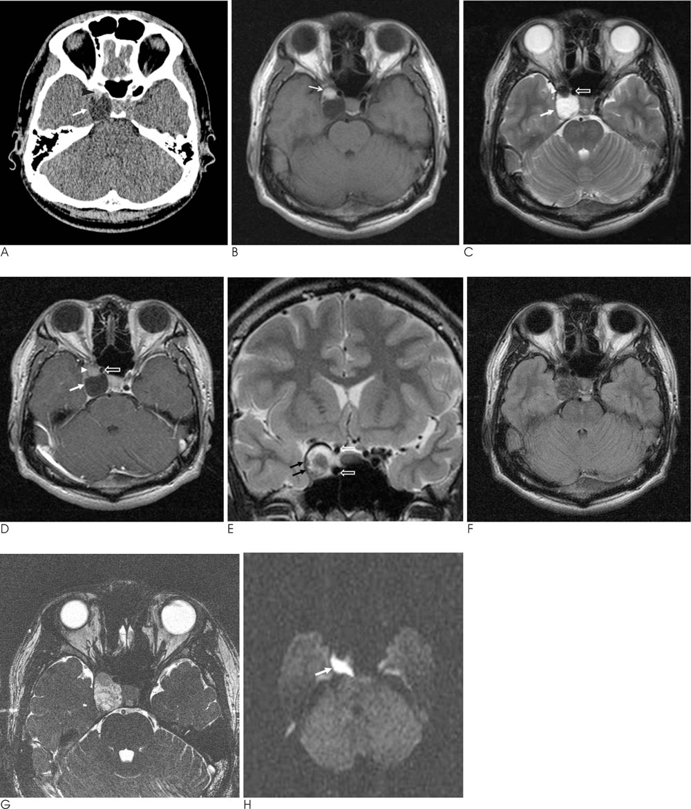
Fig. 1 A 22-years-old man with an epidermoid cyst in the right cavernous sinus. A. The precontrast CT scan demonstrated a well-defined contour-bulging oval shaped hypoattenuated mass (arrow) in the right cavernous sinus with a focal high attenuated portion (arrowhead). B-D. On the MR images, the mass (arrow) was mostly hypointense on the T1-weighted image (B), slightly heterogeneous hyperintense on the T2-weighted SE image (C) and there was no demonstrable enhancement of the mass in the right cavernous sinus on the postcontrast T1-weighted image, except for enhancement of the lateral wall of the mass (D). The anterior focal hyperintense portion (arrow) of the mass on the T1-weighted SE image was reversed in signal intensity on the T2-weighted SE image, as compared with the main cystic lesion. The cavernous segment of the right internal carotid artery (open arrows) was shifted anteromedially. E. The thickened lateral margin of the mass was seen as a dark signal rim on the T2-weighted images and as dot-like dark signal intensities (black arrows), and this represents the laterally displaced cranial nerves on the coronal T2-weighted image. F, G. The FLAIR (F) and CISS (G) sequences provide high resolution images that show the heterogenous signal intensity of the mass in the right cavernous sinus. H. The diffusion-weighted image shows a bright signal intensity mass in the right cavernous sinus (arrow).

Fig. 2 The intraoperative photograph shows the incised lateral wall of the cavernous sinus and the protruding lobulated, pearly white tumor (arrow).
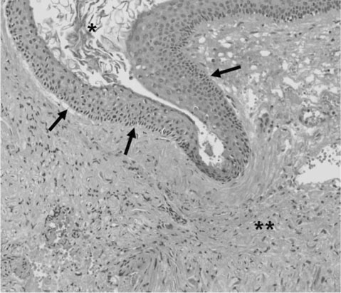
Fig. 3 Histopathological examination shows the typical epithelial lining (arrows) of the epidermoid cyst with keratin(*) and the adjacent nerve sheath cells (**) (hematoxylin and eosin stain, original magnification ×2.0).
Reference
-
1. Guidetti B, Gagliardi FM. Epidermoid and dermoid cysts. Clinical evaluation and late surgical results. J Neurosurg. 1977; 47:12–18.2. Gharabaghi A, Koerbel A, Samii A, Safavi-Abbasi S, Tatagiba M, Samii M. Epidermoid cysts of the cavernous sinus. Surg Neurol. 2005; 64:428–433.3. Osborn AG, Preece MT. Intracranial cysts: radiologic-pathologic correlation and imaging approach. Radiology. 2006; 239:650–664.4. Eekhof JL, Thomeer RT, Bots GT. Epidermoid tumor in the lateral ventricle. Surg Neurol. 1985; 23:189–192.5. Matsuno A, Takanashi S, Iwamuro H, Tanaka H, Nakaguchi H, Nagashima T. Epidermoid tumor arising in the anterior interhemispheric fissure. J Clin Neurosci. 2006; 13:262–264.6. Ikezaki K, Toda K, Abe M, Tabuchi K. Intracavernous epidermoid tumor presenting with abducens nerve paresis—case report. Neurol Med Chir (Tokyo). 1992; 32:360–364.7. Barat JL, Marchal JC, Paquis P, Auque J, Roche JL, Lepoire J. Extradural intracavernous epidermoid cyst. Apropos of 2 cases. Neurochirurgie. 1990; 36:242–245.8. Dufour H, Fuentes S, Metellus P, Grisoli F. Intracavernous epidermoid cyst. Case report and review of the literature. Neurochirurgie. 2001; 47:55–59.9. Li F, Zhu S, Liu Y, Chen G, Chi L, Qu F. Hyperdense intracranial epidermoid cysts: a study of 15 cases. Acta Neurochir (Wien). 2007; 149:31–39.10. Gao PY, Osborn AG, Smirniotopoulos JG, Harris CP. Radiologic-pathologic correlation. Epidermoid tumor of the cerebellopontine angle. AJNR Am J Neuroradiol. 1992; 13:863–872.
- Full Text Links
-
- Actions
-
Cited
- CITED
-
- Close
- Share
-
- Similar articles
-
- Useful of Three-dimensional Magnetic Rsonance Angiography and Constructive Interference in Steady State(CISS) Sequences in Patients with Hemifacial Spasm
- Diagnostic Usefulness of CISS Image in Preoperative Evaluation of Trigeminal Neuralgia and Hemifacial Spasm
- Testicular Epidermoid Cyst on Diffusion-Weighted MR Imaging and ADC Map : A Case Report
- Evaluation of Chondromalacia in the Knee Joint using Three Dimensional Fourier Transformation Constructive Interference in Steady State(CISS)
- CISS MR Imaging Findings of Epidermoid Tumor: Comparison with Spin-Echo Images
