Spinal Cord Glioblastoma Induced by Radiation Therapy of Nasopharyngeal Rhabdomyosarcoma with MRI Findings: Case Report
- Affiliations
-
- 1Department of Radiology, Seoul National University College of Medicine, Seoul 110-744, Korea. kimio@snu.ac.kr
- KMID: 1392947
- DOI: http://doi.org/10.3348/kjr.2012.13.5.652
Abstract
- Radiation-induced spinal cord gliomas are extremely rare. Since the first case was reported in 1980, only six additional cases have been reported.; The radiation-induced gliomas were related to the treatment of Hodgkin's lymphoma, thyroid cancer, and medullomyoblastoma, and to multiple chest fluoroscopic examinations in pulmonary tuberculosis patient. We report a case of radiation-induced spinal cord glioblastoma developed in a 17-year-old girl after a 13-year latency period following radiotherapy for nasopharyngeal rhabdomyosarcoma. MRI findings of our case are described.
Keyword
MeSH Terms
-
Contrast Media/diagnostic use
Female
Gadolinium DTPA/diagnostic use
Glioblastoma/*diagnosis/pathology/surgery
Humans
*Magnetic Resonance Imaging
Nasopharyngeal Neoplasms/*radiotherapy
Neoplasms, Radiation-Induced/*diagnosis/pathology
Rhabdomyosarcoma/*radiotherapy
Spinal Cord Neoplasms/*diagnosis/pathology/surgery
Figure
-
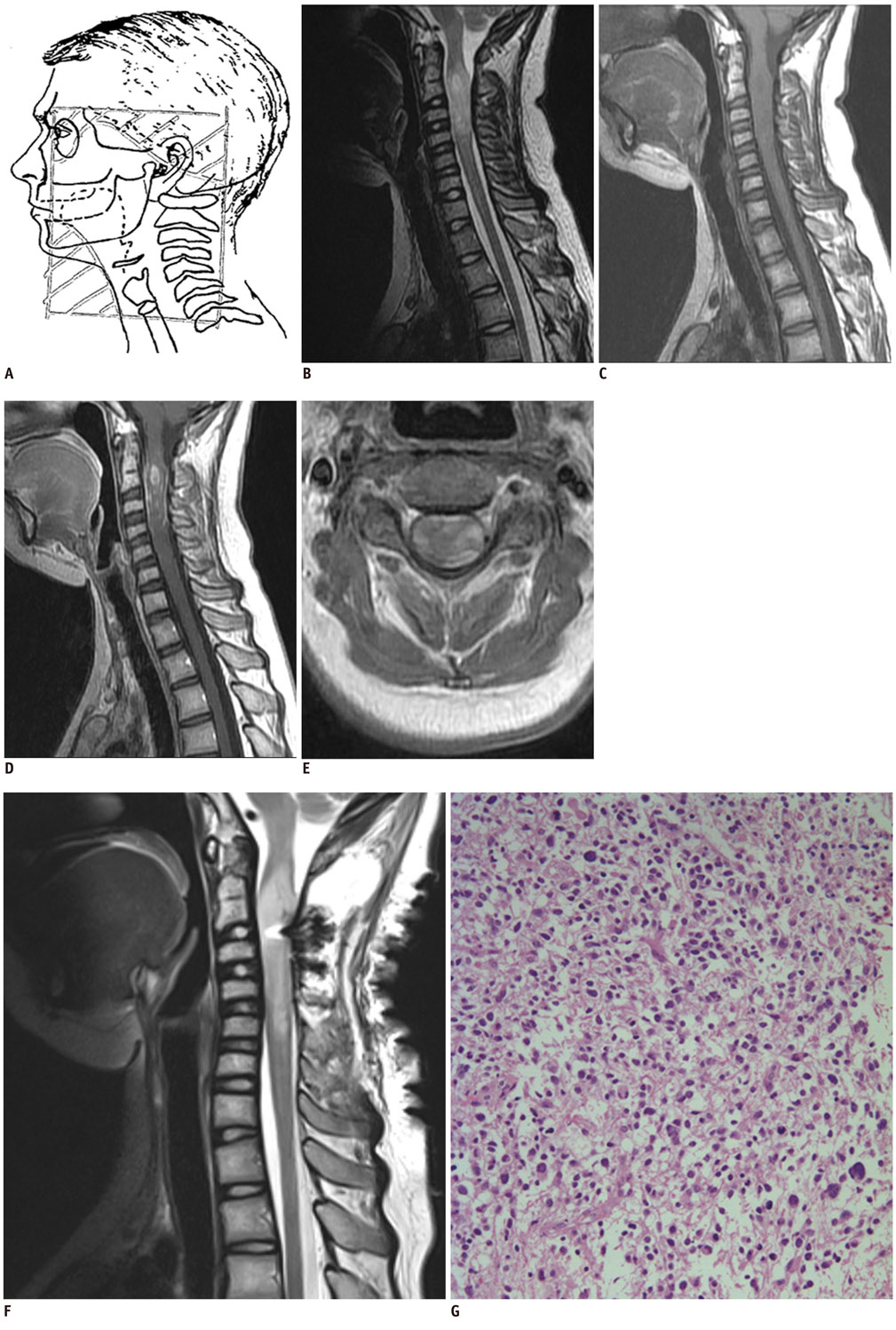
Fig. 1 Magnetic resonance imaging and pathologic findings of radiation-induced spinal cord glioblastoma. A. Radiation field. Bidirectional right and left spinal ports were used and field of radiation coverage spanned zygomatic area to C6 level. Cervical spine MRI at symptom onset. B. Sagittal T2-weighted image shows diffuse enlargement and increased signal intensity of cervical cord from C2 to C4, which is accompanied with minimal edema. C. Sagittal T1-weighted image shows iso to hypo signal intensity compared to lesion. D, E. Contrast-enhanced sagittal (D) and axial (E) T1-weighted images show ring-like enhancement at periphery, nodular discrete enhancement at upper portion of lesion, and multiple stippled foci of enhancement. F. Cervical spine MRI on 11th postsurgical day: Residual mass has become more expanded, and longitudinal extent of abnormal high signal intensity increased on sagittal T2-weighted image. G. Photomicrography of glioblastoma shows neoplastic astrocytes with high cellularity, nuclear pleomorphism, and high frequency of atypical mitosis (H & E stain, magnifications × 200).
Reference
-
1. Riffaud L, Bernard M, Lesimple T, Morandi X. Radiation-induced spinal cord glioma subsequent to treatment of Hodgkin's disease: case report and review. J Neurooncol. 2006. 76:207–211.2. Bazan C 3rd, New PZ, Kagan-Hallet KS. MRI of radiation induced spinal cord glioma. Neuroradiology. 1990. 32:331–333.3. Ng C, Fairhall J, Rathmalgoda C, Stening W, Smee R. Spinal cord glioblastoma multiforme induced by radiation after treatment for Hodgkin disease. Case report. J Neurosurg Spine. 2007. 6:364–367.4. Pettorini BL, Park YS, Caldarelli M, Massimi L, Tamburrini G, Di Rocco C. Radiation-induced brain tumours after central nervous system irradiation in childhood: a review. Childs Nerv Syst. 2008. 24:793–805.5. Steinbok P. Spinal cord glioma after multiple fluoroscopies during artificial pneumothorax treatment of pulmonary tuberculosis: case report. J Neurosurg. 1980. 52:838–841.6. Kitabatake T, Kurokawa S, Sakai K. A prospective survey on chest malignancies following multiple fluroscopies during artificial pneumothorax therapy for pulmonary tuberculosis. Tohoku J Exp Med. 1976. 118:317–322.7. Myrden JA, Hiltz JE. Breast cancer following multiple fluoroscopies during artificial pneumothorax treatment of pulmonary tuberculosis. Can Med Assoc J. 1969. 100:1032–1034.8. Howe GR. Lung cancer mortality between 1950 and 1987 after exposure to fractionated moderate-dose-rate ionizing radiation in the Canadian fluoroscopy cohort study and a comparison with lung cancer mortality in the Atomic Bomb survivors study. Radiat Res. 1995. 142:295–304.9. Calabrò F, Jinkins JR. MRI of radiation myelitis: a report of a case treated with hyperbaric oxygen. Eur Radiol. 2000. 10:1079–1084.10. Schultheiss TE, Higgins EM, El-Madhi AM. The latent period in clinical radation myelopathy. Int J Radiat Oncol Biol Phys. 1984. 10:1109–1115.11. Alfonso ER, De Gregorio MA, Mateo P, Escó R, Bascón N, Morales F, et al. Radiation myelopathy in over-irradiated patients: MR imaging findings. Eur Radiol. 1997. 7:400–404.12. Uchida K, Nakajima H, Takamura T, Kobayashi S, Tsuchida T, Okazawa H, et al. Neurological improvement associated with resolution of irradiation-induced myelopathy: serial magnetic resonance imaging and positron emission tomography findings. J Neuroimaging. 2009. 19:274–276.
- Full Text Links
-
- Actions
-
Cited
- CITED
-
- Close
- Share
-
- Similar articles
-
- Rhabdomyosarcoma of the Spermatic Cord: A Case Report
- A Case of Rhabdomyosarcoma of Spermatic Cord
- A Case of Intramedullary Glioblastoma Multiforme Involving Thoracic Cord in Child
- A Case of Spinal Cord Glioblastoma Multiforme with Intracranial Metastasis
- Primary Spinal Cord Astrocytoma Presenting as Intracranial Hypertension: A Case Report
