Ultrasonographic findings of pylorogastric intussusceptions in two dogs
- Affiliations
-
- 1College of Veterinary Medicine, Chonnam National University, Gwangju 500-757, Korea.
- 2Haemaru Referral Animal Hospital, Seongnam 463-050, Korea.
- 3College of Veterinary Medicine, Seoul National University, Seoul 151-742, Korea. heeyoon@snu.ac.kr
- KMID: 1376199
- DOI: http://doi.org/10.4142/jvs.2012.13.2.215
Abstract
- A Yorkshire terrier (case 1) and a Miniature Schnauzer (case 2) were diagnosed with pylorogastric intussusceptions (PGIs). Both cases showed acute vomiting and had previous histories of laparotomy. In case 1, the invaginated pyloric wall was thickened unevenly containing multiple hypoechoic areas and had indistinct wall layering on ultrasonography. PGI with diffuse gastric edema and necrosis was confirmed on laparotomy. The dog recovered completely after gastrectomy and a Y-U plasty. Case 2 had uniformly thickened walls of invaginated gastric pylorus with the distinct wall layering. PGI was reduced spontaneously the next day.
Keyword
MeSH Terms
Figure
-
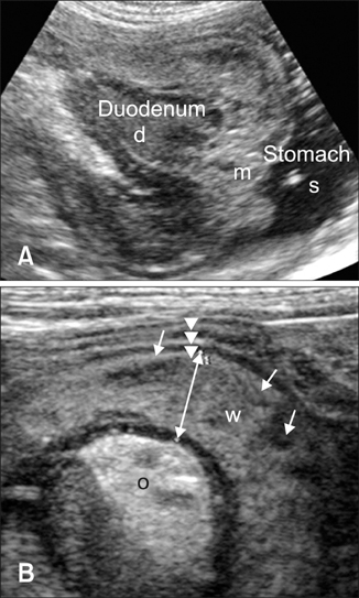
Fig. 1 Ultrasonography of a pylorogastric intussusception in case 1. (A) A mass (m), 38 × 31 mm in size, connected with the descending duodenum (d). The mass occupied the body of the stomach (s). (B) Concentric multiple rings (arrow heads) represent an intussusception. The pyloric wall (double-headed arrow and W) is thickened (7.1 mm) with indistinct layering. Short arrows are multiple hypoechoic regions in the pyloric wall. Note the edematous change of mesentery (o).
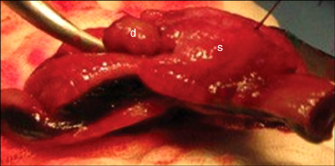
Fig. 2 Gross examination of a pylorogastric intussusception in case 1. The invaginated proximal duodenum (d), within the stomach (s).

Fig. 3 Ultrasonography a pylorogastric intussusception in case 2. (A) In the transverse view, a concentric, multiple ring (arrow) and edematous mesentery (o) was observed in the gastric body. (B) In longitudinal image of the ring, the invaginated pylorus showed thickened muscular layer (*) with distinct layering. The stomach (s) was dilated with fluid.
Reference
-
1. Applewhite AA, Cornell KK, Selcer BA. Diagnosis and treatment of intussusceptions in dogs. Compend Contin Educ Vet. 2002. 24:110–126.2. Applewhite AA, Cornell KK, Selcer BA. Pylorogastric intussusception in the dog: A case report and literature review. J Am Anim Hosp Assoc. 2001. 37:238–243.
Article3. Bowersox TS, Caywood DD, Hayden DW. Idiopathic, duodenogastric intussusception in an adult dog. J Am Vet Med Assoc. 1991. 199:1608–1609.4. Bright RM, Richardson DC, Stanton ME. Y-U antral flap advancement pyloroplasty in dogs. Compend Contin Educ Vet. 1988. 10:139–144.5. Huml RA, Konde LJ, Sellon RK, Forrest LJ. Gastrogastric intussusception in a dog. Vet Radiol Ultrasound. 1992. 33:150–153.
Article6. Lee H, Yeon S, Lee H, Chang D, Eom K, Yoon J, Choi H, Lee Y. Ultrasonographic diagnosis-pylorogastric intussusceptions in a dog. Vet Radiol Ultrasound. 2005. 46:317–318.7. Marks DL. Canine pylorogastric intussusception. Vet Med Small Anim Clin. 1983. 78:677–680.8. Patsikas MN, Jakovljevic S, Moustardas N, Papazoglou LG, Kazakos GM, Dessiris AK. Ultrasonographic signs of intestinal intussusception associated with acute enteritis or gastroenteritis in 19 young dogs. J Am Anim Hosp Assoc. 2003. 39:57–66.
Article9. Shum JS, Lo SS, Ka SY, Yeung CW, Ho JT. Gastroduodenal intussusception. Abdom Imaging. 2007. 32:698–700.
Article
- Full Text Links
-
- Actions
-
Cited
- CITED
-
- Close
- Share
-
- Similar articles
-
- Ultrasonographic measurement of optic nerve sheath diameter in normal dogs
- Laparoscopic colectomy of colonic intussusceptions in adults
- Ultrasonographic diagnosis of calcifying tendinopathy of the biceps brachii in a Doberman Pinscher dog: a case report
- Ultrasonographic findings of cataract
- Recurrent Intussusceptions in Identical Twins, Visited to Emergency Department
