Recurrent Lower Gastrointestinal Bleeding from Congenital Arteriovenous Malformation in the Terminal Ileum Mimicking Intestinal Varicosis: A Case Report
- Affiliations
-
- 1Department of Internal Medicine, Liver Research Institute Seoul National University College of Medicine, Seoul, Korea. issong@snu.ac.kr
- 2Department of Surgery, Liver Research Institute Seoul National University College of Medicine, Seoul, Korea.
- 3Department of Pathology, Liver Research Institute Seoul National University College of Medicine, Seoul, Korea.
- KMID: 1127099
- DOI: http://doi.org/10.3346/jkms.2007.22.4.746
Abstract
- We report on an exceptional vascular cause of gastrointestinal hemorrhage. A 30-yr-old man was admitted because of recurrent hematochezia. Colonoscopy showed circumferential, erythematous, and nodular vascular distensions with hematocystic spots in the terminal ileum resembling varicosis and subsequent computed tomography with 3-dimensional angiographic reconstruction revealed a vascular architecture around the terminal ileum. No other potential source of bleeding was identified. The patient was treated by ileocecectomy and the final diagnosis was of an arteriovenous malformation confined to the terminal ileum. He has been followedup without a further hemorrhagic episode.
Keyword
MeSH Terms
Figure
-
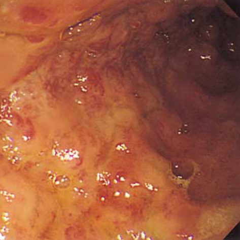
Fig. 1 Colonoscopic view of the arteriovenous malformation. This image shows circumferential, erythematous, tortuous vascular distensions with hematocystic spots in the terminal ileum, resembling the varices seen in the esophagus of patients with portal hypertension.
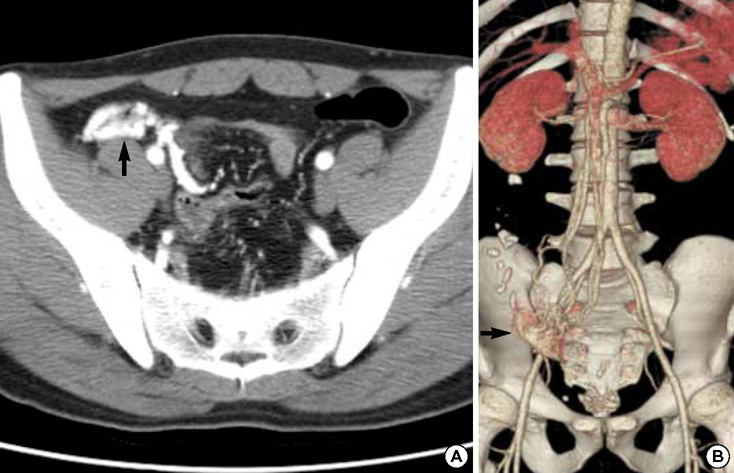
Fig. 2 Computed tomography (CT) scan finding with 3-dimensional angiographic reconstruction. The high-density architecture encasing the terminal ileum was identified on arterial phase CT (A) with delayed enhancement on venous phase. This three-dimensional angiographic reconstruction reveals a vascular tuft with a long draining vein in the right lower quadrant area (B). Arrow, arteriovenous malformation.
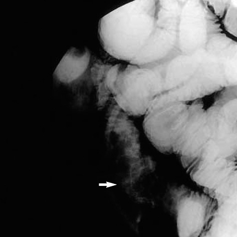
Fig. 3 Finding of enteroclysis revealed a segmental, nodular, and uneven elevated lesion with a length of 10 cm in the terminal ileum, with no other obvious source of hemorrhage in the remaining small bowel (arrow).
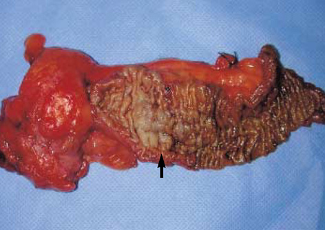
Fig. 4 Photograph of ileocecectomy specimen showing a nodular dilated vascular lesion at luminal surfaces in the terminal ileum (arrow).

Fig. 5 Photomicrograph of a pathological specimen revealing dilated thick-walled vascular structures involving all layers of the bowel wall (hematoxylin-eosin, original magnification×12.5). Arrowheads, arteriovenous malformation.
Cited by 1 articles
-
An Arteriovenous Malformation in the Jejunum Mimicking a Gastrointestinal Stromal Tumor
Eun Jeong Gong, Do Hoon Kim, Hwoon-Yong Jung, Kee Don Choi, Ho June Song, Gin Hyug Lee, Jin Ho Kim, Ho-Seop Park
Korean J Gastroenterol. 2014;63(1):42-46. doi: 10.4166/kjg.2014.63.1.42.
Reference
-
1. Meyer CT, Troncale FJ, Galloway S, Sheahan DG. Arteriovenous malformations of the bowel: an analysis of 22 cases and a review of the literature. Medicine (Baltimore). 1981. 60:36–48.2. Tang SJ, Zanati S, Dubcenco E, Cirocco M, Christodoulou D, Kandel G, Haber GB, Kortan P, Marcon NE. Diagnosis of small-bowel varices by capsule endoscopy. Gastrointest Endosc. 2004. 60:129–135.
Article3. Tang SJ, Jutabha R, Jensen DM. Push enteroscopy for recurrent gastrointestinal hemorrhage due to jejunal anastomotic varices: a case report and review of the literature. Endoscopy. 2002. 34:735–737.
Article4. Chung YW, Jeon YC, Paik CH, Kim JP, Han DS, Sohn JH, Oh YH, Park YW, Hahm JS. Pedunculated angiodysplasia of the colon treated with endoscopic resection: a case report. Dig Dis Sci. 2005. 50:1550–1552.
Article5. Nasseri-Moghaddam S, Mohamadnejad M, Malekzadeh R, Tavangar SM. Images of interest. Gastrointestinal: polypoid arteriovenous malformation of the colon. J Gastroenterol Hepatol. 2004. 19:1419.6. Maeng L, Choi KY, Lee A, Kang CS, Kim KM. Polypoid arteriovenous malformation of colon mimicking inflammatory fibroid polyp. J Gastroenterol. 2004. 39:575–578.
Article7. Ichikawa T, Koyama A, Fujimoto H, Honma M, Saiga T, Matsubara N, Ozeki Y, Uchiyama G, Ohtomo K. Diffuse arteriovenous malformation involving jejunum and total colon with mesenteric varices. Clin Imaging. 1994. 18:221–223.
Article8. Defreyne L, Meersschaut V, van Damme S, Berrevoet F, Robberecht E, Praet M. Colonic arteriovenous malformation in a child misinterpreted as an idiopathic colonic varicosis on angiography: remarks on current classification of childhood intestinal vascular malformations. Eur Radiol. 2003. 13:Suppl 4. L138–L141.
Article9. Steffani KD, Eisenberger CF, Gocht A, Izbicki JR, Yekebas EF. Recurrent intestinal bleeding in a patient with arterio-venous fistulas in the small bowel, limited mesenteric varicosis without portal hypertension and malrotation type I. Z Gastroenterol. 2003. 41:587–590.
Article10. Haringsma J, Tytgat GN. Chronic intestinal bleeding caused by congenital arteriovenous malformations. Endoscopy. 1988. 20:330–331.
Article11. Park KH, Kim YI, Yoo YO, Park SH, Lee HI, Joo DH, Kim HG, Sung NK. A case of bleeding vascular malformation of the jejunum. J Korean Surg Soc. 1998. 54:748–751.12. Moore JD, Thompson NW, Appelman HD, Foley D. Arteriovenous malformations of the gastrointestinal tract. Arch Surg. 1976. 111:381–389.
Article13. Mindelzun RE, Beaulieu CF. Using biphasic CT to reveal gastrointestinal arteriovenous malformations. Am J Roentgenol. 1997. 168:437–438.
Article
- Full Text Links
-
- Actions
-
Cited
- CITED
-
- Close
- Share
-
- Similar articles
-
- Arteriovenous Malformation of the Distal Ileum in a 14-Year-Old Girl with Recurrent Abdominal Pain: A Case Report
- A Case of Arteriovenous Malformation of the Descending Colon Presenting with Massive Bleeding
- A Case of Successful Transarterial Embolization in Arteriovenous Malformation of Uterus
- A Case of Arteriovenous Malformation of the Pancreas
- A Case of Lower Gastrointestinal Bleeding due to Angiodysplasia in the Terminal Ileum
