Recurrent Massive Subcutaneous Hemorrhage in Neurofibromatosis Type 1: A Case Report
- Affiliations
-
- 1Department of Emergency Medicine, College of Medicine, Inha University, Incheon, Korea. LIFSAV@inha.ac.kr
- 2Department of Chest Surgery, College of Medicine, Inha University, Incheon, Korea.
- 3Department of Pathology, College of Medicine, Inha University, Incheon, Korea.
- KMID: 1127095
- DOI: http://doi.org/10.3346/jkms.2007.22.4.728
Abstract
- Neurofibromatosis type 1 (NF-1) is an autosomal dominant disorder that has three major features: multiple neural tumors, cafe-au-lait spots, and pigmented iris hamartomas (Lisch nodules). The purpose of this case report is to advise physicians of the danger associated with the progression of fast-onset massive hemorrhage to hemodynamic instability, which mandates rapid treatment to prevent the development of a life-threatening condition. A 64-yr-old woman with NF-1 was admitted to the Emergency Department (ED) because of a rapidly growing, 10x5x3 cm-sized mass on the left back area. She had previously undergone surgery for a large subcutaneous hematoma, which had developed on her right back area 30 yr before. She became hemodynamically unstable with hypotension during the next 3 hr after admission to ED. Resuscitation and blood transfusion were done, and the hematoma was surgically removed. The mass presented as a subcutaneous, massive hematoma with pathologic findings of neurofibroma. We report a case of NF-1 that presented as recurrent, massive, subcutaneous hemorrhage on the back region combined with hypovolemic shock.
Keyword
MeSH Terms
Figure
-
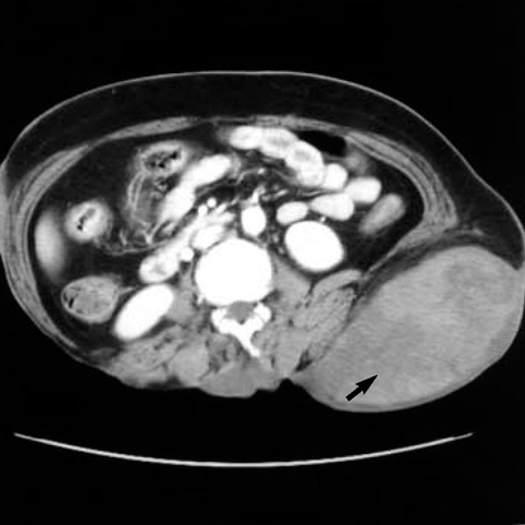
Fig. 1 Abdomen and pelvis CT showing a huge, subcutaneous, 13×7.6 cm-sized hematoma, along with fluid collection in the posterior and left lateral subcutaneous layer of the thoracolumbar and left lower back area (arrow).
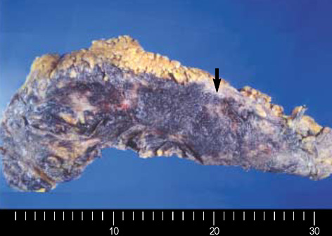
Fig. 2 The tumor is ill-defined, pale yellow, soft, and fibrotic and spreads into the fat tissue (arrow). Extensive hemorrhage is present on the surface of, but not inside, the tumor.

Fig. 3 The tumor is composed of interlacing bundles of elongated cells with wavy nuclei with the fibrillary collagenous background and shows infiltrative growth into the fat tissue (H&E, ×100).
Cited by 1 articles
-
Transcatheter Embolization of a Ruptured Internal Pudendal Artery Pseudoaneurysm in a Patient with Neurofibromatosis Type 1
Chang-Wei Zhang, Zhi-Gang Yang, Xiao-Dong Xie, Chao-Hua Wang, Chao You, Wei Li
J Korean Med Sci. 2010;25(4):638-640. doi: 10.3346/jkms.2010.25.4.638.
Reference
-
1. Rao V, Affifi RA, Ghazarian D. Massive subcutaneous hemorrhage in a chest-wall neurofibroma. Can J Surg. 2000. 43:459–460.2. Poston GJ, Grace PA, Venn G, Spencer J. Recurrence near-fatal haemorrhage in von Recklinghausen's disease. Br J Clin Pract. 1990. 44:755–756.3. White N, Gwanmesia I, Akhtar N, Withey SJ. Severe haemorrhage in neurofibroma: a lesson. Br J Plast Surg. 2004. 57:456–457.4. Restrepo CS, Riascos RF, Hatta AA, Rojas R. Neurofibromatosis type 1 spinal manifestations of a systemic disease. J Comput Assist Tomogr. 2005. 29:532–539.5. Tsutsumi M, Kazekawa K, Tanaka A, Ueno Y, Nomoto Y, Nii K, Haraoka S. Rapid expansion of benign scalp neurofibroma caused by massive intramural hemorrhage. Neurol Med Chir (Tokyo). 2002. 42:338–340.6. Lin YC, Chen HC. Rare complication of massive hemorrhage in neurofibromatosis with arteriovenous malformation. Ann Plast Surg. 2000. 44:221–224.
Article7. Gottfried ON, Viskochil DH, Fults DW, Couldwell WT. Molecular, genetic, and cellular pathogenesis of neurofibromas and surgical implications. Neurosurgery. 2006. 58:1–16.
Article
- Full Text Links
-
- Actions
-
Cited
- CITED
-
- Close
- Share
-
- Similar articles
-
- Spontaneous Massive Hemothorax in a Patient with Neurofibromatosis Type 1 with Successful Transarterial Embolization
- Recurrent Shoulder Dislocation and Proximal Humerus Fracture in Neurofibromatosis Type I: A Case Report
- Spontaneous Hemothorax Caused by Rupture of an Intercostal Artery Aneurysm in Neurofibromatosis Type I I : A Case Report
- A Case of Orbital Neurilemoma Associated with Neurofibroma tosis
- Spontaneous Hemothorax in a Patient with Type I Neurofibromatosis
