Decreased Left Ventricular Torsion and Untwisting in Children with Dilated Cardiomyopathy
- Affiliations
-
- 1Department of Pediatrics, Seoul National University Children's Hospital, Seoul National University College of Medicine, SeoulKorea. ksydhnoh@yahoo.co.kr
- 2Department of Pediatrics, Eulji Medical Center, Eulji University, Seoul, Korea.
- KMID: 1127078
- DOI: http://doi.org/10.3346/jkms.2007.22.4.633
Abstract
- The purpose of this study was to analyze left ventricular (LV) torsion and untwisting, and to evaluate the correlation between torsion and other components of LV contraction in children with dilated cardiomyopathy (DCM). Segmental and global rotation, rotational rate (Vrot) were measured at three levels of LV using the twodimensional (2D) speckle tracking imaging (STI) method in 10 DCM patients (range 0.6-15 yr, median 6.5 yr, 3 females) and 17 age- and sex-matched normal controls. Global torsion was decreased in DCM (peak global torsion; 10.9+/-4.6degrees vs. 0.3+/-2.1degrees, p<0.001). Loss of LV torsion occurred mainly by the diminution of counterclockwise apical rotation and was augmented by somewhat less reduction in clockwise basal rotation. In DCM, the normal counterclockwise apical rotation was not observed, and the apical rotation about the central axis was clockwise or slightly counterclockwise (peak apical rotation; 5.9+/-4.1degrees vs. -0.9+/-3.1degrees, p<0.001). Systolic counterclockwise Vrot and early diastolic clockwise Vrot at the apical level were decreased or abolished. In DCM, decreased systolic torsion and loss of early diastolic recoil contribute to LV systolic and diastolic dysfunction. The STI method may facilitate the serial evaluation of the LV torsional behavior in clinical settings and give new biomechanical concepts for better management of patients with DCM.
Keyword
MeSH Terms
Figure
-
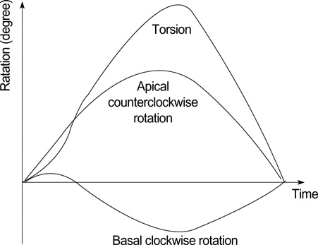
Fig. 1 Measurement of the torsion (degree). Torsion (t)=Apical LV rotation (t)-Basal LV rotation (t).

Fig. 2 (A) Profiles of apical segmental and global (dotted line) rotation (°) in a 6 month-old normal boy. Apical rotation was consistently counterclockwise (positive). (B) Profiles of apical segmental and global (dotted line) rotation (°) in a 6 month-old boy with DCM. Apical rotation was clockwise or small counterclockwise, and was markedly heterogenous.
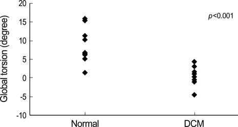
Fig. 3 Comparison of peak global torsion in normal and DCM children. The peak global torsion was significantly decreased in DCM.
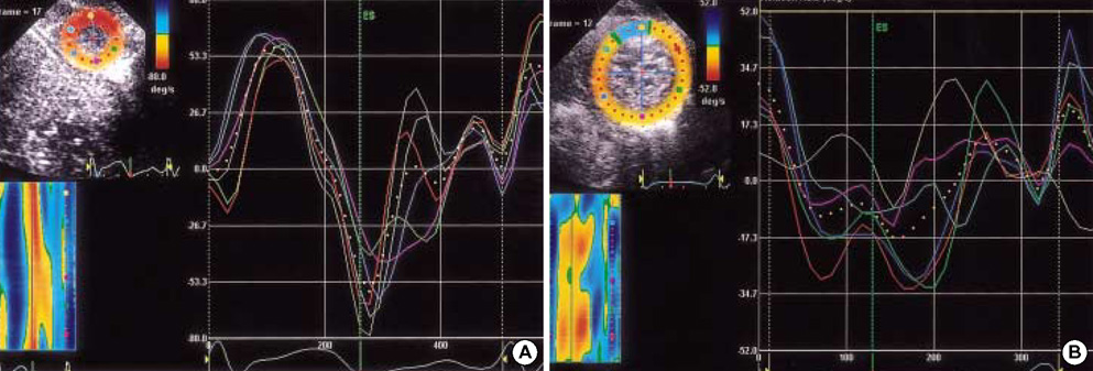
Fig. 4 (A) Profiles of apical segmental and global (dotted line) rotational rate (Vrot) (°/sec) in a 6 month-old normal boy. LV apex showed systolic counterclockwise (positive) Vrot and clockwise early diastolic (negative) Vrot. (B) Profiles of apical segmental and global (dotted line) rotational rate (Vrot) (°/sec) in a 6 month-old boy with DCM. Systolic counterclockwise Vrot and early diastolic clockwise Vrot were abolished. Considerable rotational heterogeneity was noticed particularly at the apical level.
Reference
-
1. Buckberg GD, Clemente C, Cox JL, Coghlan HC, Castella M, Torrent-Guasp F, Gharib M. The structure and function of the helical heart and its buttress wrapping. IV. Concepts of dynamic function from the normal macroscopic helical structure. Semin Thorac Cardiovasc Surg. 2001. 13:342–357.
Article2. Burleson KO, Schwartz GE. Cardiac torsion and electromagnetic fields: the cardiac bioinformation hypothesis. Medical Hypotheses. 2005. 64:1109–1116.
Article3. Yun KL, Niczyporuk MA, Daughters GT, Ingels NB Jr, Stinson EB, Alderman EL, Hansen DE, Miller DC. Alterations in left ventricular diastolic twist mechanics during acute human cardiac allograft rejection. Circulation. 1991. 83:962–973.
Article4. Notomi Y, Srinath G, Shiota T, Martin-Miklovic MG, Beachler L, Howell K, Oryszak SJ, Deserranno DG, Freed AD, Greenberg NL, Younoszai A, Thomas JD. Maturational and adaptive modulation of left ventricular torsional biomechanics: Doppler tissue imaging observation from infancy to adulthood. Circulation. 2006. 113:2534–2541.5. Tibayan FA, Lai DT, Timek TA, Dagum P, Liang D, Daughters GT, Ingels NB, Miller DC. Alterations in left ventricular torsion in tachycardia-induced dilated cardiomyopathy. J Thorac Cardiovasc Surg. 2002. 124:43–49.
Article6. Setser RM, Kasper JM, Lieber ML, Starling RC, McCarthy PM, White RD. Persistent abnormal left ventricular systolic torsion in dilated cardiomyopathy after partial left ventriculectomy. J Thorac Cardiovasc Surg. 2003. 126:48–55.
Article7. Suhling M, Jansen C, Arigovindan M, Buser P, Marsch S, Unser M, Hunziker P. Multiscale motion mapping; a novel computer vision technique for quantitative, objective echocardiographic motion measurement independent of Doppler; first clinical description and validation. Circulation. 2004. 110:3093–3099.8. Helak JW, Reichek N. Quantification of human left ventricular mass and volume by 2-dimensional echocardiography: in vivo anatomic validation. Circulation. 1981. 63:1398–1407.9. Sutton M, Plappert T, Spigel A, Raichlen J, Douglas P, Reichek N, Edmunds L. Early prospective changes in left ventricular chamber size, architecture, and function in aortic stenosis and aortic regurgitation and their relation to intraoperative changes in afterload: a prospective 2-dimensional echocardiographic study. Circulation. 1987. 76:77–89.10. Notomi Y, Lysyansky P, Setser RM, Shiota T, Popovic JB, Martin-Miklovic MG, Weaver JA, Oryszak SJ, Greenberg NL, White RD, Thomas JD. Measurement of ventricular torsion by two-dimensional ultrasound speckle tracking imaging. J Am Coll Cardiol. 2005. 45:2034–2041.
Article11. Notomi Y, Setser RM, Shiota T, Martin-Miklovic MG, Weaver JA, Popovic JB, Yamada H, Greenberg NL, White RD, Thomas JD. Assessment of left ventricular torsional deformation by Doppler tissue imaging: validation study with tagged magnetic resonance imaging. Circulation. 2005. 111:1141–1147.12. Becker M, Bilke E, Kühl H, Katoh M, Kramann R, Franke A, Bucker A, Hanrath P, Hoffmann R. Analysis of myocardial deformation based on pixel tracking in 2D echocardio-graphic images allows quantitative assessment of regional left ventricular function. Heart. 2006. 92:1102–1108.13. Toyoda T, Bada H, Akasaka T, Akiyama M, Neishi Y, Tomita J, Sukmawan R, Koyama Y, Watanabe N, Tamano S, Shinomura R, Komuro I, Yoshida K. Assessment of regional myocardial strain by a novel automated tracking system from digital image files. J Am Soc Echocardiogr. 2004. 17:1234–1238.
Article14. Yun KL, Miller DC. Torsional deformation of the left ventricle. J Heart Valve Dis. 1995. 4:214–222.15. LeGrice IJ, Smail BH, Chai LZ, Edgar SG, Gavin JB, Hunter PJ. Laminar structure of the heart: ventricular myocyte arrangement and connective tissue architecture in the dog. Am J Physiol. 1995. 269:H571–H582.
Article16. Arts T, Costa KD, Covell JW, McCulloch AD. Relating myocardial laminar structure to shear strain and muscle fiber orientation. Am J Physiol Circ Physiol. 2001. 280:2222–2229.17. Spotnitz HM. Macro design, structure, and mechanics of the left ventricle. J Thorac Cardiovasc Surg. 2000. 119:1053–1077.
Article18. Young AA, Dokos S, Powell KA, Sturm B, McCulloch AD, Starling RC, McCarthy PM, White RD. Regional heterogeneity of function in nonischemic dilated cardiomyopathy. Cardiovasc Res. 2001. 49:308–318.
Article19. Notomi Y, Martin-Miklovic MG, Oryszak SJ, Shiota T, Deserranno D, Popovic ZB, Garcia MJ, Greenberg NL, Thomas JD. Enhanced ventricular untwisting during exercise: a mechanistic manifestation of elastic recoil described by Doppler tissue imaging. Circulation. 2006. 113:2524–2533.20. Bach DS, Beanlands RS, Schwaiger M, Armstrong WF. Heterogeneity of ventricular function and myocardial oxidative metabolism in nonischemic dilated cardiomyopathy. J Am Coll Cardiol. 1995. 25:1258–1262.
Article21. Hayashida W, Kumada T, Nohara R, Tanio H, Kambayashi M, Ishikawa N, Nakamura Y, Himura Y, Kawai C. Left ventricular regional wall stress in dilated cardiomyopathy. Circulation. 1990. 82:2075–2083.
Article22. Juillière Y, Marie PY, Danchin N, Gillet C, Paille F, Karcher G, Bertrand A, Cherrier F. Radionuclide assessment of regional differences in left ventricular wall motion and myocardial perfusion in idiopathic dilated cardiomyopathy. Eur Heart J. 1993. 14:1163–1169.23. Nakayama Y, Shimizu G, Hirota Y, Saito T, Kino M, Kitaura Y, Kawamura K. Functional and histopathologic correlation in patients with dilated cardiomyopathy: an integrated evaluation by multicariate analysis. J Am Coll Cardiol. 1987. 10:186–192.24. Chow E, Woodard JC, Farrar DJ. Rapid ventricular pacing in pigs: an experimental model of congestive heart failure. Am J Physiol. 1990. 258:H1603–H1605.
Article25. Spinale FG, Zellner JL, Johnson WS, Eble DM, Munyer PD. Cellular and extracellular remodeling with the development and recovery from tachycardia-induced cardiomyopathy: changes in fibrillar collagen, myocyte adhesion capacity and proteoglycans. J Mol Cell Cardiol. 1996. 28:1591–1608.
Article26. Spinale FG, Tomita M, Zellner JL, Cook JC, Crawford FA, Zile MR. Collagen remodeling and changes in LV funcition during development and recovery from supraventricular tachycardia. Am J Physiol. 1991. 261:H308–H318.27. Spinale FG, Coker ML, Thomas CV, Walker JD, Mukherjee R, Hebbar L. Time-dependent changes in matrix metalloproteinase activity and expression during the progression of congestive heart failure: relation to ventricular and myocyte function. Circ Res. 1998. 82:482–495.28. Delhaas T, Arts T, Prinzen FW, Reneman RS. Regional electrical activation and mechanical function in the partially ischemic left ventricle of dogs. Am J Physiol. 1996. 271:H2411–H2420.
Article29. O'Rourke B, Kass DA, Tomaselli GF, Kaab S, Tunin R, Marban E. Mechanisms of altered excitation-contraction coupling in canine tachycardia-induced heart failure, I: experimental studies. Circ Res. 1999. 84:562–570.30. Winslow RL, Rice J, Jafri S, Marban E, O'Rourke B. Mechanisms of altered excitation-contraction coupling in canine tachycardia-induced heart failure, II: model studies. Circ Res. 1999. 84:571–586.31. Torrent-Guasp F, Kocica MJ, Corno AF, Komeda M, Cox J, Flotats A, Ballester-Rodes M, Carreras-Costa F. Systolic ventricular filling. Eur J Cardiothorac Surg. 2004. 25:376–386.32. Yip G, Wang M, Zhang Y, Fung JW, Ho PY, Sanderson JE. Left ventricular long axis function in diastolic heart failure is reduced in both diastole and systole: time for redefinition? Heart. 2002. 87:121–125.33. Henein MJ, Gibson DG. Normal long-axis function. Heart. 1999. 81:111–113.34. Henein MJ, Gibson DG. Long-axis function in disease. Heart. 1999. 81:229–231.
- Full Text Links
-
- Actions
-
Cited
- CITED
-
- Close
- Share
-
- Similar articles
-
- Hemodynamics and Left Ventricular Cineangiographic Findings in Idiopathic Dilated Cardiomyopathy
- Vanishing Left Ventricular Thrombi in Severe Aortic Stenosis with Dilated Cardiomyopathy
- A Study of Left Ventricular Function by Digitized Echocardiograms in Dilated Cardiomyopathy
- Left Ventricular Dysfunction and Dilated Cardiomyopathy in Infants and Children with Wolff-Parkinson-White Syndrome in the Absence of Tachyarrhythmias
- A Case of Normalized Hypertrophic Cardiomyopathy after Removal of Pheochromocytoma
