Solitary Small Hepatic Angiosarcoma: Initial and Follow-up Imaging Findings
- Affiliations
-
- 1Department of Diagnostic Radiology, Chonnam National University Hwasun Hospital, Hwasun, Korea. yjeong@chonnam.ac.kr
- 2Department of Diagnostic Radiology, Chonnam National University Hospital, Gwangju, Korea.
- KMID: 1126844
- DOI: http://doi.org/10.3348/kjr.2007.8.2.180
Abstract
- We report an uncommon case of solitary, small hepatic angiosarcoma that was initially considered as a hemangioma. We present the imaging findings, with an emphasis on the initial and follow-up CT and MR findings, as well as report on the more suggestive findings of angiosarcoma than those of a hemangioma.
MeSH Terms
Figure
-
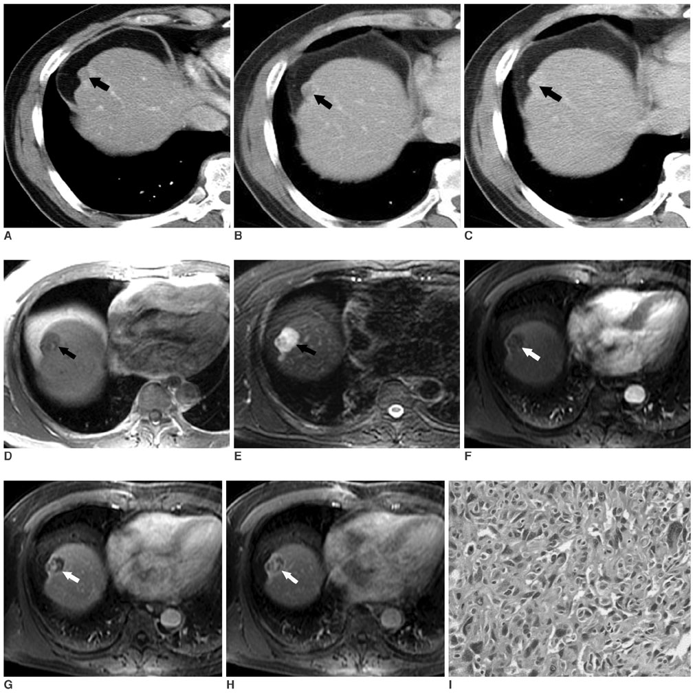
Fig. 1 A 60-year-old man with hepatic angiosarcoma. A. The arterial phase contrast-enhanced CT image shows a 1.1 cm low attenuated lesion in the hepatic dome (arrow). B. The portal-venous phase contrast-enhanced CT image shows some peripheral nodular enhancement (arrow). C. The delayed phase contrast-enhanced CT image shows a persistent, homogeneous enhancing mass (arrow). D. The T1-weighted gradient-echo (TR/TE = 120/4.2 msec) MR image three months later shows a hypointense mass with focal hyperintense foci that were considered as hemorrhage within the mass in the hepatic dome. Note the increased size of the mass (arrow) compared with the initial CT (A-C). E. The fat-suppressed T2-weighted fast spin-echo (TR/TE = 6666/91 msec) MR image shows a heterogeneous hyperintense mass with areas of low signal intensity (arrow). F. The arterial phase contrast-enhanced gradient echo (TR/TE = 180/1.5 msec) MR image shows faint peripheral rim enhancement (arrow). G. The portal-venous phase contrast-enhanced gradient echo (TR/TE = 180/1.5 msec) MR image reveals centrally septal-like enhancement (arrow). H. The delayed phase contrast-enhanced gradient echo (TR/TE = 180/1.5 msec) MR image shows heterogeneous and persistent enhancement (arrow). I. Photograph (original magnification, ×400; H & E staining) of the specimen shows the numerous and irregular vascular spaces separated by fibrous septa and lined by pleomorphic and hyperchromatic endothelial tumor cells.
Cited by 1 articles
-
Clinical Courses of Primary Hepatic Angiosarcoma: Retrospective Analysis of Eight Cases
Chang Jae Hur, Bo Ram Min, Yoo Jin Lee, Byung Kuk Jang, Jae Seok Hwang, Eun Soo Kim, Kyung Sik Park, Kwang Bum Cho, Yu Na Kang, Woo Jin Chung
Korean J Gastroenterol. 2015;65(4):229-235. doi: 10.4166/kjg.2015.65.4.229.
Reference
-
1. Buetow PC, Buck JL, Ros PR, Goodman ZD. Malignant vascular tumors of the liver: radiologic-pathologic correlation. Radiographics. 1994. 14:153–166.2. Koyama T, Fletcher JG, Johnson CD, Kuo MS, Notohara K, Burgart LJ. Primary hepatic angiosarcoma: findings at CT and MR imaging. Radiology. 2002. 222:667–673.3. Peterson MS, Baron RL, Rankin SC. Hepatic angiosarcoma: findings on multiphasic contrast-enhanced helical CT do not mimic hepatic hemangioma. AJR Am J Roentgenol. 2000. 175:165–170.4. Itai Y, Teraoka T. Angiosarcoma of the liver mimicking cavernous hemangioma on dynamic CT. J Comput Assist Tomogr. 1989. 13:910–912.5. Ohmoto K, Hirokawa M, Takesue M, Yamamoto S. Hepatic angiosarcoma with early central enhancement and arterioportal shunt on dynamic CT. Hepatogastroenterology. 2000. 47:1717–1718.6. Jones EC, Chezmar JL, Nelson RC, Bernardino ME. The frequency and significance of small (less than or equal to 15 mm) hepatic lesions detected by CT. AJR Am J Roentgenol. 1992. 158:535–539.7. Robinson PJ, Arnold P, Wilson D. Small "indeterminate" lesions on CT of the liver: a follow-up study of stability. Br J Radiol. 2003. 76:866–874.8. Blachar A, Federle MP, Brancatelli G. Hepatic capsular retraction: spectrum of benign and malignant etiologies. Abdom Imaging. 2002. 27:690–699.
- Full Text Links
-
- Actions
-
Cited
- CITED
-
- Close
- Share
-
- Similar articles
-
- Primary Colonic Epithelioid Angiosarcoma with Hepatic Metastasis: A Case Report
- Hepatic and splenic angiosarcoma: A case report
- Hepatic Angiosarcoma Mimicking Cavernous Hemangioma on Dynamic CT
- Atypical Angiosarcoma with a Solitary Erythematous Nodule on the Cheek: A Case Report
- A Case Report of Breast Angiosarcoma in a Young Woman
