Actinomycosis of the Gallbladder Mimicking Carcinoma: a Case Report with US and CT Findings
- Affiliations
-
- 1Department of Radiology, Wonju Christian Hospital, Wonju College of Medicine, Yonsei University, Wonju, Korea. kshyun@smc.samsung.co.kr
- 2Department of Radiology and Center for Imaging Science, Samsung Medical Center, Sungkyunkwan University School of Medicine, Seoul, Korea.
- 3Department of Pathology, Wonju Christian Hospital, Wonju College of Medicine, Yonsei University, Wonju, Korea.
- 4Department of Surgery, Wonju Christian Hospital, Wonju College of Medicine, Yonsei University, Wonju, Korea.
- KMID: 1126841
- DOI: http://doi.org/10.3348/kjr.2007.8.2.169
Abstract
- We describe a case of actinomycosis of the gallbladder mimicking carcinoma. Sonography showed a hypoechoic mass replacing gallbladder lumen and engulfing a stone; contrast-enhanced computed tomography showed a heterogeneously enhanced thickened gallbladder wall with subtle, disrupted luminal surface enhancement, which formed a mass. As a result of the clinical and radiologic presentation, our impression was of gallbladder carcinoma. Actinomycosis should be included in the differential diagnosis when sonography and computed tomography findings show a mass engulfing the stone in the gallbladder and extensive pericholecystic infiltration with extension to neighboring abdominal wall muscle.
Keyword
MeSH Terms
Figure
-
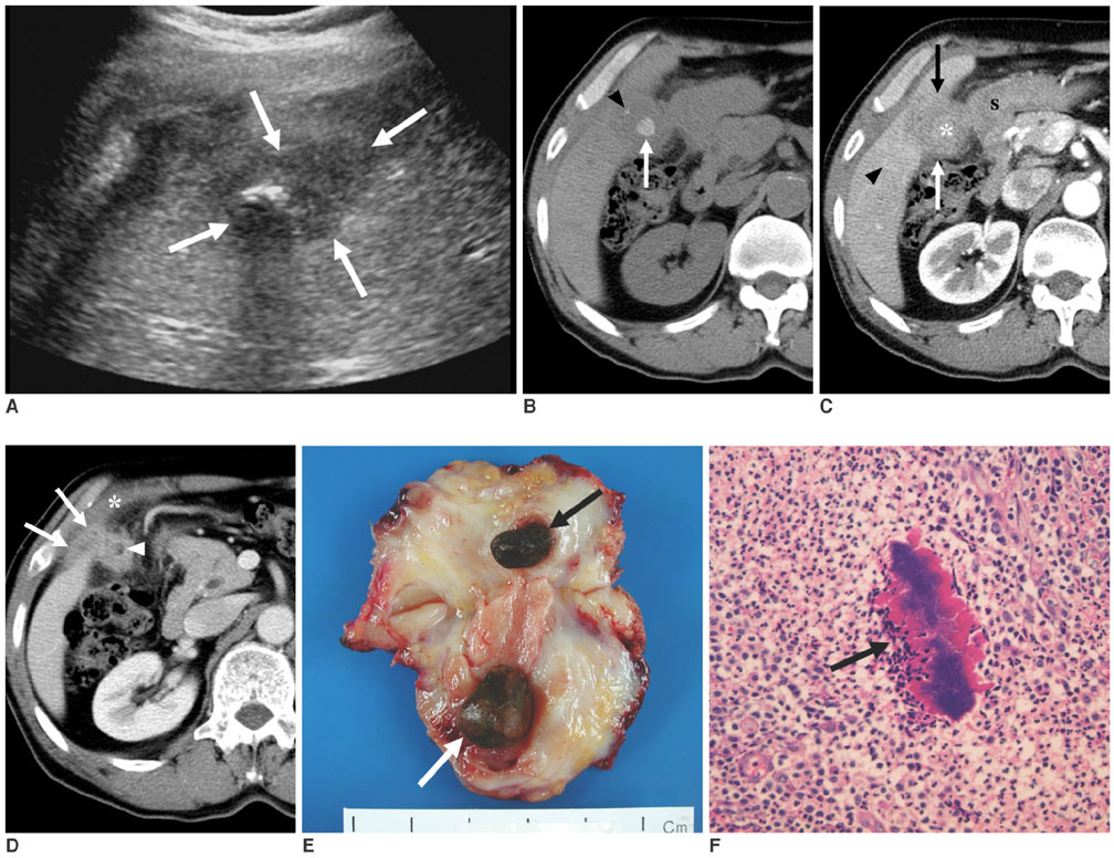
Fig. 1 A 65-year-old man with actinomycosis of the gallbladder which presented as a mass. A. Sonograph shows a hypoechoic mass (arrows) replacing gallbladder lumen and engulfing a gallstone with infiltration of surrounding liver. B. Unenhanced CT scan shows the impacted gallstone (arrow) and calcification (arrowhead) in the thickened gallbladder wall. The other gallstone is in the neck of the gallbladder (not shown). C. Contrast-enhanced CT scan obtained during the arterial phase at the same level as B, shows a heterogeneously enhanced and markedly thickened gallbladder wall (arrows) with infiltration of surrounding liver, stomach, and pericholecystic fatty tissue. Faint, disrupted luminal surface enhancement around the stone (asterisk) and hepatic parenchymal enhancement (arrowhead) adjacent to the thickened gallbladder wall are evident. S = stomach. D. Contrast-enhanced CT scan obtained during the portal phase at a level 1 cm below C shows disrupted luminal surface enhancement of the thickened gallbladder wall (arrowhead) with pericholecystic fatty infiltration, perihepatic extension of the soft tissue mass (arrows), and mild thickening of the transversus abdominis muscle (asterisk). E. Photograph of the cut surface of the resected gallbladder shows markedly thickening of gallbladder body and fundus walls and a fibrotic appearance. Two brown stones (arrows) are indicated. F. Photomicrograph of the thickened gallbladder wall shows inflammatory cell infiltration with fibrosis, and a sulfur granule (arrow) containing tangled filamentous bacilli, which is compatible with Actinomyces. (H & E stain, × 400).
Reference
-
1. Lee IJ, Ha HK, Park CM, Kim JK, Kim JH, Kim TK, et al. Abdominopelvic actinomycosis involving the gastrointestinal tract: CT features. Radiology. 2001. 220:76–80.2. Merle-Melet M, Mory F, Stempfel B, Maurer P, Regent D, Parent S, et al. Actinomyces naeslundii, acute cholecystitis, and carcinoma of the gallbladder. Am J Gastroenterol. 1995. 90:1530–1531.3. Marrie T, Stiver HG, Molgat A, Stark RG, Norris D. Actinomycosis of the gallbladder. Can J Surg. 1977. 20:147–149.4. Brewer JH, Allen MJ. Actinomycosis of the gallbladder with liver abscess. South Med J. 1980. 73:1070–1072.5. Freland C, Massoubre B, Horeau JM, Caillon J, Drugeon HB. Actinomycosis of the gallbladder due to Actinomyces naeslundii. J Infect. 1987. 15:251–257.6. van Steensel CJ, Kwan TS. Actinomycosis of the gallbladder. Neth J Surg. 1988. 40:23–25.7. Ormsby AH, Bauer TW, Hall GS. Actinomycosis of the cholecystic duct: case report and review. Pathology. 1998. 30:65–67.8. Nakayama F. Recent progress in the diagnosis and treatment of carcinoma of the gallbladder--introduction. World J Surg. 1991. 15:313–314.9. Chun KA, Ha HK, Yu ES, Shinn KS, Kim KW, Lee DH, et al. Xanthogranulomatous cholecystitis: CT features with emphasis on differentiation from gallbladder carcinoma. Radiology. 1997. 203:93–97.10. Wilbur AC, Sagireddy PB, Aizenstein RI. Carcinoma of the gallbladder: color Doppler ultrasound and CT findings. Abdom Imaging. 1997. 22:187–189.11. Das DK, Tripathi RP, Bhambhani S, Chachra KL, Sodhani P, Malhotra V. Ultrasound-guided fine-needle aspiration cytology diagnosis of gallbladder lesions: a study of 82 cases. Diagn Cytopathol. 1998. 18:258–264.12. Kazmierski RH. Primary adenocarcinoma of the gallbladder with intramural calcification. Am J Surg. 1951. 82:248–250.13. Cintron JR, Del Pino A, Duarte B, Wood D. Abdominal actinomycosis. Dis Colon Rectum. 1996. 39:105–108.14. Schlech WF 3rd, Gelfand M, Alper B, Kaiser AB. Medical management of visceral actinomycosis. South Med J. 1983. 76:921–922.
