Recurrent Bilateral Branch Retinal Artery Occlusion with Hearing Loss and Encephalopathy: The First Case Report of Susac Syndrome in Korea
- Affiliations
-
- 1Department of Ophthalmology, Asan Medical Center, University of Ulsan, College of Medicine, Seoul, Korea. yhyoon@amc.seoul.kr
- 2Department of Neurology, Asan Medical Center, University of Ulsan, College of Medicine, Seoul, Korea.
- 3Department of Radiology, Asan Medical Center, University of Ulsan, College of Medicine, Seoul, Korea.
- 4Department of Otolaryngology, Asan Medical Center, University of Ulsan, College of Medicine, Seoul, Korea.
- KMID: 1123456
- DOI: http://doi.org/10.3346/jkms.2011.26.11.1518
Abstract
- We report the first case of Susac syndrome in Koreans, in a 23-yr-old female patient who presented with sudden visual loss and associated neurological symptoms. Ophthalmic examination and fluorescein angiography showed multiple areas of branch retinal artery occlusion, which tended to recur in both eyes. Magnetic resonance imaging showed dot-like, diffusion-restricted lesions in the corpus callosum and left fornix, and audiometry showed low-frequency sensory hearing loss, compatible with Susac syndrome. She received immunosuppressive therapy with oral steroid and azathioprine. Three months later all the symptoms disappeared but obstructive vasculitis have been relapsing. This patient demonstrated the entire clinical triad of Susac syndrome, which tends to occur in young females. Although this disorder has rarely been reported in Asian populations, a high index of suspicion is warranted for early diagnosis and timely treatment.
MeSH Terms
-
Autoimmune Diseases/diagnosis/drug therapy
Azathioprine/administration & dosage/*therapeutic use
Brain/blood supply/pathology
Female
Hearing Loss
Humans
Immunotherapy
Magnetic Resonance Imaging
Republic of Korea
Retinal Artery Occlusion/diagnosis/drug therapy/pathology
Susac Syndrome/*diagnosis/*drug therapy/pathology
Young Adult
Figure
-

Fig. 1 Fundus photo showing edematous lesions in the supra-temporal area (A). Fluorescein angiography, showing a hyperfluorescent arterial wall proximal to the obstructed branch retinal artery (B).
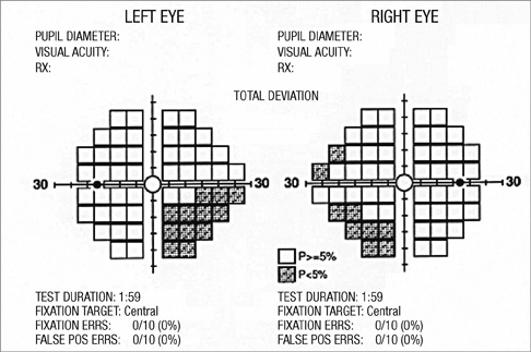
Fig. 2 Visual field test results, showing an infranasal field defect of the left eye corresponding to the obstructed lesion and a small area of visual field defect in the right eye.
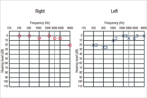
Fig. 3 Pure tone audiometry, showing decreased sensitivity to low-frequency sounds in the left ear.
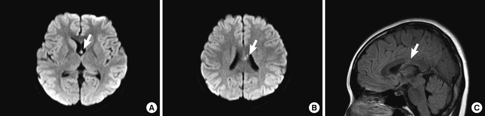
Fig. 4 Magnetic resonance image of the brain. (A, B) Diffusion weighted images, showing a diffusion restricted lesion in the corpus callosum and left fornix. (C) Sagittal Fluid-attenuated inversion recovery (FLAIR) image showing a focal signal change in the corpus callosum.
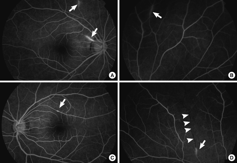
Fig. 5 Fluorescein angiography showing hyperfluorescent vasculitis lesions (arrows) (A-D) and an obstructed branch retinal artery (arrowheads) (D).
Reference
-
1. Murata Y, Inada K, Negi A. Susac syndrome. Am J Ophthalmol. 2000. 129:682–684.2. Rennebohm RM, Egan RA, Susac JO. Treatment of Susac's syndrome. Curr Treat Options Neurol. 2008. 10:67–74.3. Susac JO, Egan RA, Rennebohm RM, Lubow M. Susac's syndrome: 1975-2005 microangiopathy/autoimmune endotheliopathy. J Neurol Sci. 2007. 257:270–272.4. Rennebohm R, Susac JO, Egan RA, Daroff RB. Susac's syndrome: update. J Neurol Sci. 2010. 299:86–91.5. Jarius S, Neumayer B, Wandinger KP, Hartmann M, Wildemann B. Anti-endothelial serum antibodies in a patient with Susac's syndrome. J Neurol Sci. 2009. 285:259–261.6. Martinet N, Fardeau C, Adam R, Bodaghi B, Papo T, Piette JC, Lehoang P. Fluorescein and indocyanine green angiographies in Susac syndrome. Retina. 2007. 27:1238–1242.7. Trevino R, Pearlman R. Idiopathic recurrent branch retinal arterial occlusion in a young adult. Optom Vis Sci. 1998. 75:11–16.8. Beatty S, Au Eong KG. Acute occlusion of the retinal arteries: current concepts and recent advances in diagnosis and management. J Accid Emerg Med. 2000. 17:324–329.9. Recchia FM, Brown GC. Systemic disorders associated with retinal vascular occlusion. Curr Opin Ophthalmol. 2000. 11:462–467.10. Gass JD, Tiedeman J, Thomas MA. Idiopathic recurrent branch retinal arterial occlusion. Ophthalmology. 1986. 93:1148–1157.11. Notis CM, Kitei RA, Cafferty MS, Odel JG, Mitchell JP. Microangiopathy of brain, retina, and inner ear. J Neuroophthalmol. 1995. 15:1–8.12. Susac JO, Hardman JM, Selhorst JB. Microangiopathy of the brain and retina. Neurology. 1979. 29:313–316.13. O'Halloran HS, Pearson PA, Lee WB, Susac JO, Berger JR. Microangiopathy of the brain, retina, and cochlea (Susac syndrome). A report of five cases and a review of the literature. Ophthalmology. 1998. 105:1038–1044.14. Susac JO. Susac's syndrome: the triad of microangiopathy of the brain and retina with hearing loss in young women. Neurology. 1994. 44:591–593.15. Dörr J, Radbruch H, Bock M, Wuerfel J, Bruggemann A, Wandinger KP, Zeise D, Pfueller CF, Zipp F, Paul F. Encephalopathy, visual disturbance and hearing loss-recognizing the symptoms of Susac syndrome. Nat Rev Neurol. 2009. 5:683–688.
- Full Text Links
-
- Actions
-
Cited
- CITED
-
- Close
- Share
-
- Similar articles
-
- A Case of Susac Syndrome
- Bilateral Hearing Loss in Wernicke Encephalopathy
- Clinical observation of the bilateral branch vein occlusion
- Central Retinal Artery Occlusion Masquerading as Branch Retinal Artery Occlusion
- Vertebrobasilar Occlusion Presenting as Sudden Isolated Bilateral Sensorineural Hearing Loss: Case Report
