Individual Pulmonary Vein Atresia in Adults: Report of Two Cases
- Affiliations
-
- 1Department of Radiology, Soonchunhyang University Cheonan Hospital, Cheonan 330-721, Korea. ytokim@schca.ac.kr
- KMID: 1122339
- DOI: http://doi.org/10.3348/kjr.2011.12.3.395
Abstract
- We present two cases of individual pulmonary vein atresia without vestige of an involved pulmonary vein. On CT, we noted the absence or interruption of normal pulmonary venous structures, and the presence of abnormal vascular structures that represented collaterals for the involved lung parenchyma. On angiography, the atretic pulmonary vein was found to drain into the other ipsilateral pulmonary veins through the collaterals.
Keyword
MeSH Terms
Figure
-
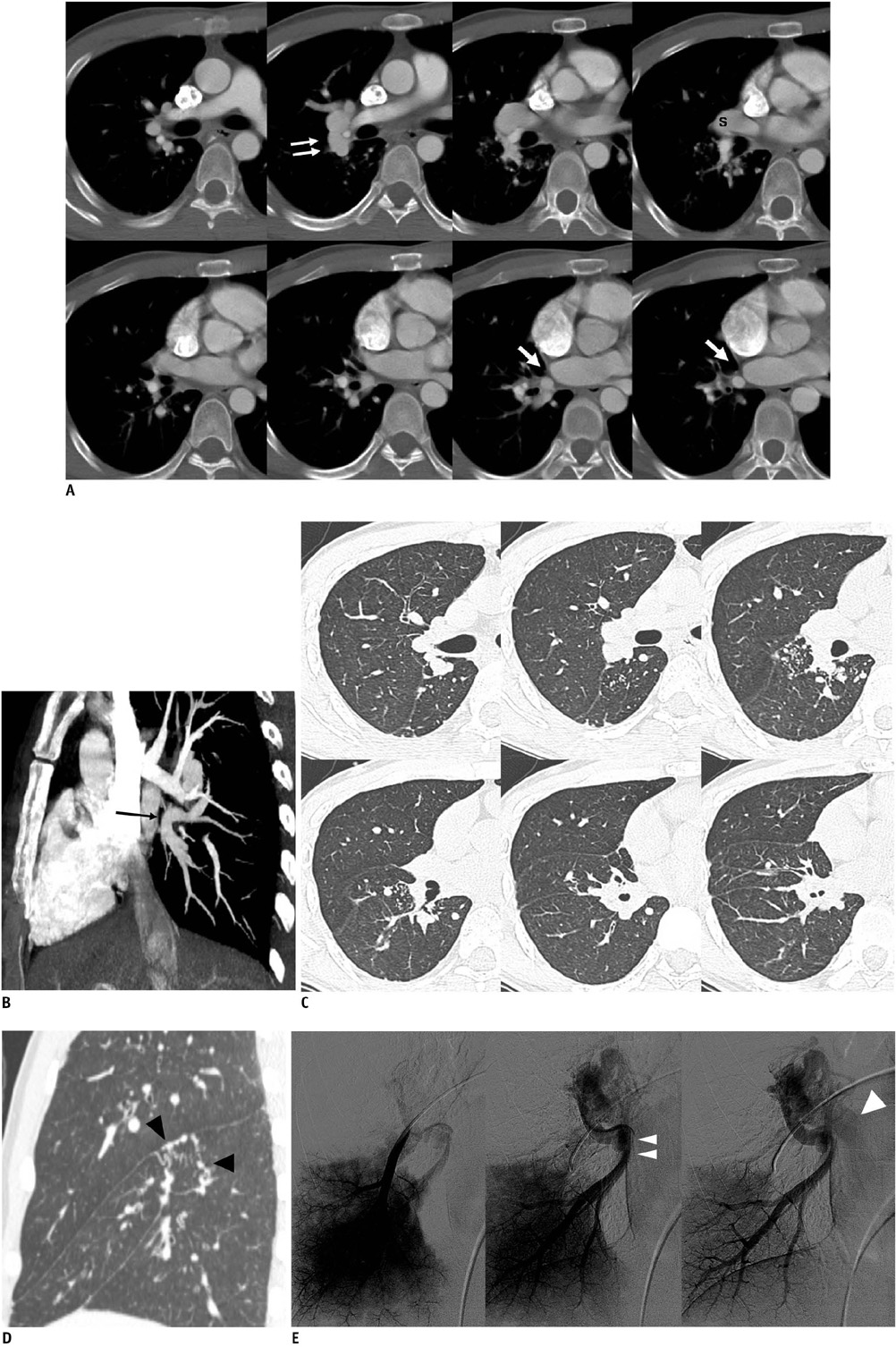
Fig. 1 Lobar pulmonary vein atresia of right lower lobe in 32-year-old man. A. Serial axial images show low-attenuation linear structure (arrows) between right inferior pulmonary vein and left atrium, as well as prominent right superior pulmonary vein(s). Right pulmonary artery is not hypoplastic compared with left pulmonary artery. Also note abnormally enlarged vascular structure (double arrows) near right pulmonary artery, suggesting presence of collateral between superior pulmonary vein and interrupted inferior pulmonary vein on angiogram. B. Reformatted oblique sagittal image shows atretic right inferior pulmonary vein (black arrow). C. Serial axial images with lung setting show multiple dot-like collaterals between superior pulmonary vein and atretic inferior pulmonary vein in right lower lobe. D. Reformatted coronal image shows tortuous and dot-like collaterals (black arrowheads) in superior segment of right lower lobe. E. Angiogram of right lower lobe shows atretic right inferior pulmonary vein (double arrowheads) draining into superior pulmonary vein (arrowhead) through collaterals.
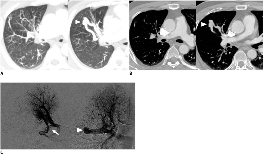
Fig. 2 Segmental pulmonary vein atresia of apical segment from right upper lobe in 43-year-old man. A. Slap axial images with lung setting show tortuous and dot-like collaterals in anterior segment of right upper lobe. In addition, abnormal tubular vascular structure (arrowhead) is present in anterior segment of right upper lobe. This finding is consistent with imaging findings of pulmonary arteriovenous malformation, but we diagnosed it as collateral, from apical segment to anterior segment of right upper lobe on pulmonary angiography. B. Serial axial images show absence of right superior pulmonary vein in normal position and presence of collaterals (arrowhead) draining into segmental pulmonary vein of right upper lobe. C. Angiogram of apical segment of right upper lobe shows that apical segmental pulmonary vein is interrupted (arrow) and drains into another segmental pulmonary vein through collaterals (arrowhead).
Reference
-
1. Heyneman LE, Nolan RL, Harrison JK, McAdams HP. Congenital unilateral pulmonary vein atresia: radiologic findings in three adult patients. AJR Am J Roentgenol. 2001. 177:681–685.2. Cullen S, Deasy PF, Tempany E, Duff DF. Isolated pulmonary vein atresia. Br Heart J. 1990. 63:350–354.3. Freedom RM, Mawson JB, Yoo S, Benson LN. Congenital heart disease: textbook of angiocardiography. vol. II. 1997. New York: Futura Publishing;665–691.4. Saida Y, Eguchi N, Mori K, Tanaka YO, Ishikawa S, Itai Y. Isolated pulmonary vein stenosis associated with full intrapulmonary compensation. AJR Am J Roentgenol. 1999. 173:961–962.5. Pourmoghadam KK, Moore JW, Khan M, Geary EM, Madan N, Wolfson BJ, et al. Congenital unilateral pulmonary venous atresia: definitive diagnosis and treatment. Pediatr Cardiol. 2003. 24:73–79.6. Swischuk LE, L'Heureux P. Unilateral pulmonary vein atresia. AJR Am J Roentgenol. 1980. 135:667–672.7. Kuhn M. Cardiac and intestinal natriuretic peptides: insights from genetically modified mice. Peptides. 2005. 26:1078–1085.8. Amplatz K, Moller JH. Radiology of congenital heart disease. 1993. St. Louis: Mosby;847–909.
- Full Text Links
-
- Actions
-
Cited
- CITED
-
- Close
- Share
-
- Similar articles
-
- Cystic Lung Changes in a Thin Section CT in an Asymptomatic Young Adult with Unilateral Pulmonary Vein Atresia: A Case Report
- Unilateral Pulmonary Vein Atresia: A Case Report
- Isolated Unilateral Pulmonary Vein Atresia
- Visualization of the Pulmonary Arteries in the Patients with Pulmonary Atresia or Hypoplasia by Pulmonary Vein Wedge Angiography
- Tricuspid atresia: a re-evaluation and classification
