Optimization of the Contrast Mixture Ratio for Simultaneous Direct MR and CT Arthrography: an in Vitro Study
- Affiliations
-
- 1Department of Radiology and Institute of Radiation Medicine, Seoul National University College of Medicine, Seoul, Korea.
- 2Department of Radiology, Seoul National University Bundang Hospital, Gyeongi-do, Korea. kanghs@radcom.snu.ac.kr
- KMID: 1118873
- DOI: http://doi.org/10.3348/kjr.2008.9.6.520
Abstract
OBJECTIVE
This study was designed to determine the optimal mixture ratio of gadolinium and iodinated contrast agent for simultaneous direct MR arthrography and CT arthrography. MATERIALS AND METHODS: An in vitro study was performed utilizing mixtures of gadolinium at six different concentrations (0.625, 1.25, 2.5, 5.0, 10 and 20 mmol/L) and iodinated contrast agent at seven different concentrations (0, 12.5, 25, 37.5, 50, 75 and 92-99.9%). These mixtures were placed in tissue culture plates, and were then imaged with CT and MR (with T1-weighted sequences, proton-density sequences and T2-weighted sequences). CT numbers and signal intensities were measured. Pearson's correlation coefficients were used to assess the correlations between the gadolinium/iodinated contrast agent mixtures and the CT numbers/MR signal intensities. Scatter diagrams were plotted for all gadolinium/iodinated contrast agent combinations and two radiologists in consensus identified the mixtures that yielded the optimal CT numbers and MR signal intensities. RESULTS: The CT numbers showed significant correlation with iodinated contrast concentrations (r = 0.976, p < 0.001), whereas the signal intensities as measured on MR images showed a significant correlation with both gadolinium and iodinated contrast agent concentrations (r = -484 to -0.719, p < 0.001). A review of the CT and MR images, graphs, and scatter diagram of 42 combinations of the contrast agent showed that a concentration of 1.25 mmol/L gadolinium and 25% iodinated contrast agent was the best combination for simultaneous CT and MR imaging. CONCLUSION: A mixture of 1.25 mmol/L gadolinium and 25% iodinated contrast agent was found to be optimal for simultaneous direct MR arthrography and CT arthrography.
Keyword
MeSH Terms
-
*Arthrography
Contrast Media/*administration & dosage
Gadolinium/administration & dosage/*diagnostic use
Iohexol/administration & dosage/*analogs & derivatives/diagnostic use
*Magnetic Resonance Imaging
Meglumine/administration & dosage/*diagnostic use
Organometallic Compounds/administration & dosage/*diagnostic use
Phantoms, Imaging
*Tomography, X-Ray Computed
Figure
-
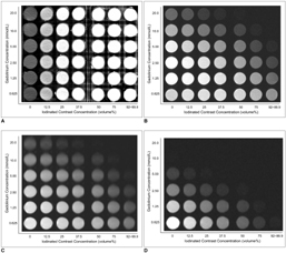
Fig. 1 CT and MR images of tissue plates used in present study. A. CT image. B. T1-weighted MR image (TR/TE = 500/14). C. Proton density MR image (TR/TE = 3000/14, ETL = 5). D. T2-weighted MR image (TR/TE = 3000/98, ETL = 12). ETL = echo train length
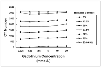
Fig. 2 Changes in CT numbers (region of interest values) at various gadolinium concentrations with different volume percentages of iodinated contrast agent.
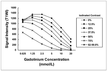
Fig. 3 Changes in signal intensities (region of interest values) at various gadolinium concentrations with different volume percentages of iodinated contrast agent as seen on proton density MR images (TR/TE = 3000/14, ETL = 5). ETL = echo train length

Fig. 4 Changes in signal intensities (region of interest values) at various gadolinium concentrations with different volume percentages of iodinated contrast agent as seen on T1-weighted MR images (TR/TE = 500/14).
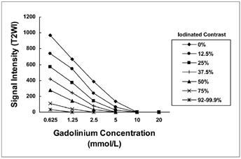
Fig. 5 Changes in signal intensities (region of interest values) at different gadolinium concentrations with different volume percentages of iodinated contrast agent as seen on T2-weighted MR images (TR/TE = 3000/98, ETL = 12). ETL = echo train length
Cited by 1 articles
-
Correlation of Structural Bony Abnormalities and Mechanical Symptoms of Hip Joints
Sung-Hwa Lyu, Yoon-Ho Kwak, Young-Kyun Lee, Yong-Chan Ha, Kyung-Hoi Koo
Hip Pelvis. 2014;26(2):115-123. doi: 10.5371/hp.2014.26.2.115.
Reference
-
1. Hajek PC, Baker LL, Sartoris DJ, Neumann CH, Resnick D. MR arthrography: anatomic-pathologic investigation. Radiology. 1987. 163:141–147.2. Czerny C, Hofmann S, Neuhold A, Tschauner C, Engel A, Recht MP, et al. Lesions of the acetabular labrum: accuracy of MR imaging and MR arthrography in detection and staging. Radiology. 1996. 200:225–230.3. Montgomery DD, Morrison WB, Schweitzer ME, Weishaupt D, Dougherty L. Effects of iodinated contrast and field strength on gadolinium enhancement: implications for direct MR arthrography. J Magn Reson Imaging. 2002. 15:334–343.4. Sciulli RL, Boutin RD, Brown RR, Nguyen KD, Muhle C, Lektrakul N, et al. Evaluation of the postoperative meniscus of the knee: a study comparing conventional arthrography, conventional MR imaging, MR arthrography with iodinated contrast material, and MR arthrography with gadolinium-based contrast material. Skeletal Radiol. 1999. 28:508–514.5. Zanetti M, Hodler J. Contrast media in MR arthrography of the glenohumeral joint: intra-articular gadopentetate vs saline: preliminary results. Eur Radiol. 1997. 7:498–502.6. Farber JM. CT arthrography and postoperative musculoskeletal imaging with multichannel computed tomography. Semin Musculoskelet Radiol. 2004. 8:157–166.7. Schmid MR, Pfirrmann CW, Hodler J, Vienne P, Zanetti M. Cartilage lesions in the ankle joint: comparison of MR arthrography and CT arthrography. Skeletal Radiol. 2003. 32:259–265.8. Vande Berg BC, Lecouvet FE, Poilvache P, Jamart J, Materne R, Lengele B, et al. Assessment of knee cartilage in cadavers with dual-detector spiral CT arthrography and MR imaging. Radiology. 2002. 222:430–436.9. Lecouvet FE, Simoni P, Koutaissoff S, Vande Berg BC, Malghem J, Dubuc JE. Multidetector spiral CT arthrography of the shoulder. Clinical applications and limits, with MR arthrography and arthroscopic correlations. Eur J Radiol. 2008. 68:120–136.10. Schmid MR, Schertler T, Pfirrmann CW, Saupe N, Manestar M, Wildermuth S, et al. Interosseous ligament tears of the wrist: comparison of multi-detector row CT arthrography and MR imaging. Radiology. 2005. 237:1008–1013.11. Kopka L, Funke M, Fischer U, Keating D, Oestmann J, Grabbe E. MR arthrography of the shoulder with gadopentetate dimeglumine: influence of concentration, iodinated contrast material, and time on signal intensity. AJR Am J Roentgenol. 1994. 163:621–623.12. Masi JN, Newitt D, Sell CA, Daldrup-Link H, Steinbach L, Majumdar S, et al. Optimization of gadodiamide concentration for MR arthrography at 3 T. AJR Am J Roentgenol. 2005. 184:1754–1761.13. Blum AG, Simon JM, Cotten A, Quirin-Cosmidis I, Boyer B, Boutry N, et al. Comparison of double-contrast CT arthrography image quality with nonionic contrast agents: isotonic dimeric iodixanol 270 mg I/mL and monomeric iohexol 300 mg I/mL. Invest Radiol. 2000. 35:304–310.14. Engel A. Magnetic resonance knee arthrography. Enhanced contrast by gadolinium complex in the rabbit and in humans. Acta Orthop Scand Suppl. 1990. 240:1–57.15. Jinkins JR, Robinson JW, Sisk L, Fullerton GD, Williams RF. Proton relaxation enhancement associated with iodinated contrast agents in MR imaging of the CNS. AJNR Am J Neuroradiol. 1992. 13:19–27.
- Full Text Links
-
- Actions
-
Cited
- CITED
-
- Close
- Share
-
- Similar articles
-
- Feasibility of Ultrasound-Guided Intraarticular Contrast Injection for MR Arthrography
- Ankle Ligaments: Comparison of MR Arthrography with Conventional MR Imaging in Amputated Feet
- Double contrast CT arthrographic findings of shoulder instability
- Assessment of anterior shoulder instability by CT arthrography
- Feasibility of Ultrasonography and MR Arthrography during Evaluation of Rotator Cuff Injury
