Small Malignant Hepatic Tumor Detection in Gadolinium- and Ferucarbotran-Enhanced Magnetic Resonance Imaging: does Combining Ferucarbotran-Enhanced T2*-Weighted Gradient Echo and T2-Weighted Turbo Spin Echo Images have Additive Efficacy?
- Affiliations
-
- 1Department of Diagnostic Radiology, Chonbuk National University Hospital and Medical School, Choubuk, Korea. jmyr@dreamwiz.com
- 2Department of Diagnostic Radiology, Wonkwang University Hospital and School of Medicine, Iksan, Korea.
- KMID: 1118872
- DOI: http://doi.org/10.3348/kjr.2008.9.6.510
Abstract
OBJECTIVE
To determine if a combination of ferucarbotran-enhanced T2*weighted-gradient echo (T2*W-GRE) and T2-weighted turbo spin echo (T2W-TSE) images in gadolinium- and ferucarbotran-enhanced MRI has additive efficacy compared to each image alone for detecting small (< or = 2.0 cm) hepatocellular carcinoma (HCC) lesions in a group of cirrhotic patients and metastases in a group of non-cirrhotic patients. MATERIALS AND METHODS: Two readers retrospectively analyzed gadolinium- and ferucarbotran-enhanced T2*W-GRE, T2W-TSE, and combined T2*W-GRE/T2W-TSE images of 119 patients with 157 HCCs and 32 patients with 98 metastases. The diagnostic accuracy and sensitivity for each image set and the combined set were evaluated using the alternative-free response receiver operating characteristic method. RESULTS: The mean area under the curve value of the combined set (0.966) tended to be better than that for each individual image set (T2W-TSE [0.910], T2*W-GRE [0.892]). Sensitivities in the combined set were higher than those in each individual image set for detecting HCC (mean, 93.0% versus 81.6% and 86.7%, respectively, p < 0.01). Sensitivities in the combined set and the T2W-TSE set were the same for detecting metastases, and both were higher than the sensitivity seen in the T2*W-GRE set (mean, 97.5% versus 85.2 %, p < 0.01). CONCLUSION: Combining ferucarbotran-enhanced T2*W-GRE and T2W-TSE has additive efficacy for detecting HCC in cirrhotic patients, but T2W-TSE is preferred for detecting metastases in non-cirrhotic patients.
MeSH Terms
-
Adult
Aged
Carcinoma, Hepatocellular/*diagnosis
Contrast Media/*administration & dosage
Female
Gadolinium DTPA/*diagnostic use
Humans
Iron/*diagnostic use
Liver Neoplasms/*diagnosis/secondary
*Magnetic Resonance Imaging/methods
Male
Middle Aged
Observer Variation
Oxides/*diagnostic use
Predictive Value of Tests
Sensitivity and Specificity
Figure
-

Fig. 1 55-year-old man with well-differentiated hepatocellular carcinoma in subcapsular area of right hepatic lobe. A. Axial respiratory-triggered T2-weighted turbo spin echo (4200/76) imaging shows no hepatic mass. B. Axial arterial phase three-dimensional dynamic MRI after administration of gadopentetate dimeglumine (4.3/2.0) shows no detected lesion. C. Axial ferucarbotran-enhanced breath-hold T2*-weighted gradient echo imaging (180/12) shows high signal intensity nodule (arrow) in subcapsular area of liver, which was assigned confidence level of 4 by both observers. D. Axial ferucarbotran-enhanced respiratory-triggered T2-weighted turbo spin echo imaging (4200/76) shows no definitive lesion (arrow) at same location as in A. This was assigned score of 1 and 2 by the two observers, respectively.

Fig. 2 50-year-old man with two hepatocellular carcinomas in right hepatic lobe. A. Axial arterial phase three-dimensional dynamic MRI after administration of gadopentetate dimeglumine (4.3/2.0) shows subtle small nodular enhancement (arrow) in subcapsular area of right hepatic lobe. Inferior margin of main mass located at slightly higher level is shown (small arrows). B. Axial ferucarbotran-enhanced breath-hold T2*-weighted gradient echo imaging (180/12) shows only main mass (arrows) in posterior portion of right hepatic lobe. Subcapsular lesion was assigned score of 1 by both observers using T2*-weighted gradient echo image set. C. Axial ferucarbotran-enhanced respiratory-triggered T2-weighted turbo spin echo imaging (4200/76) shows definitive round hyperintense subcapsular mass (arrow) and main mass (small arrows), which were assigned confidence level of 4 by both observers.
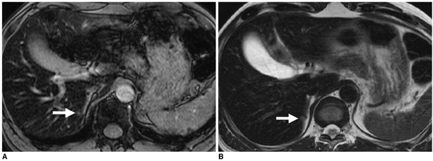
Fig. 3 57-year-old man with surgically confirmed 0.4-cm liver metastasis from rectal cancer. A. Axial ferucarbotran-enhanced breath-hold T2*-weighted gradient echo imaging (180/12) shows irregular high signal intensity lesion (arrow) at medial margin of right hepatic lobe, which was missed by one observer and was assigned confidence level of 2 by other observer during image interpretation. B. Axial ferucarbotran-enhanced respiratory-triggered T2-weighted turbo spin echo imaging (4200/76) clearly shows small round hyperintense mass (arrow) at same location as in A. This was assigned score of 4 by both observers.

Fig. 4 55-year-old man with surgically confirmed 0.4-cm liver metastasis from colon cancer. A. Axial ferucarbotran-enhanced breath-hold T2*-weighted gradient echo imaging (180/12) shows irregular high signal intensity lesion (arrow) in inferior portion of right hepatic lobe. Both reviewers missed this lesion during image interpretation. B. Axial ferucarbotran-enhanced respiratory-triggered T2-weighted turbo spin echo imaging (4200/76) clearly shows small round hyperintense mass (arrow) at same location as in A. This was assigned score of 3 by both observers.
Reference
-
1. Vogl TJ, Pegios W, McMahon C, Balzer J, Waitzinger J, Pirovano G, et al. Gadobenate dimeglumine - a new contrast agent for MR imaging: preliminary evaluation in healthy volunteers. AJR Am J Roentgenol. 1992. 158:887–892.2. Lee JM, Kim IH, Kwak HS, Youk JH, Han YM, Kim CS. Detection of small hypervascular hepatocellular carcinomas in cirrhotic patients: comparison of superparamagnetic iron oxide-enhanced MR imaging with dual-phase spiral CT. Korean J Radiol. 2003. 4:1–8.3. Tanimoto A, Wakabayashi G, Shinmoto H, Nakatsuka S, Okuda S, Kuribayashi S. Superparamagnetic iron oxide-enhanced MR imaging for focal hepatic lesions: a comparison with CT during arterioportography plus CT during hepatic arteriography. J Gastroenterol. 2005. 40:371–380.4. Kim MJ, Kim JH, Chung JJ, Park MS, Lim JS, Oh YT. Focal hepatic lesions: detection and characterization with combination gadolinium - and superparamagnetic iron oxide-enhanced MR imaging. Radiology. 2003. 228:719–726.5. Ward J, Guthrie JA, Scott DJ, Atchley J, Wilson D, Davies MH, et al. Hepatocellular carcinoma in the cirrhotic liver: double-contrast MR imaging for diagnosis. Radiology. 2000. 216:154–162.6. Seneterre E, Taourel P, Bouvier Y, Pradel J, Van Beers B, Daures JP, et al. Detection of hepatic metastases: ferumoxides-enhanced MR imaging versus unenhanced MR imaging and CT during arterial portography. Radiology. 1996. 200:785–792.7. Fretz CJ, Elizondo G, Weissleder R, Hahn PF, Stark DD, Ferrucci JT Jr. Superparamagnetic iron oxide-enhanced MR imaging: pulse sequence optimization for detection of liver cancer. Radiology. 1989. 172:393–397.8. Van Beers BE, Lacrosse M, Jamart J, Grandin C, Gigot JF, Horsmans Y, et al. Detection and segmental location of malignant hepatic tumors: comparison of ferumoxides-enhanced gradient-echo and T2-weighted spin-echo MR imaging. AJR Am J Roentgenol. 1997. 168:713–717.9. Kim SH, Choi D, Lim JH, Lee WJ, Jang HJ, Lim HK, et al. Optimal pulse sequence for ferumoxides-enhanced MR imaging used in the detection of hepatocellular carcinoma: a comparative study using seven pulse sequences. Korean J Radiol. 2002. 3:87–97.10. Ward J, Chen F, Guthrie JA, Wilson D, Lodge JP, Wyatt JI, et al. Hepatic lesions detection after superparamagnetic iron oxide enhancement: comparison of five T2-weighted sequences at 1.0 T by using alternative-free response receiver operating characteristic analysis. Radiology. 2000. 214:159–166.11. Edmondson HA, Steiner PE. Primary carcinoma of liver: study of 100 cases among 48,900 necropsies. Cancer. 1954. 7:462–503.12. Lencioni R, Pinto F, Armillotta N, Di Giulio M, Gaeta P, Di Candio G, et al. Intrahepatic metastatic nodules of hepatocellular carcinoma detected at lipiodol CT: imaging-pathologic correlation. Abdom Imaging. 1997. 22:253–258.13. Kim MJ, Kim JH, Choi JY, Park SH, Chung JJ, Kim KW, et al. Optimal TE for SPIO-enhanced gradient-recalled echo MRI for the detection of focal hepatic lesions. AJR Am J Roentgenol. 2006. 187:W255–W266.14. Chakraborty DP, Winter LH. Free-response methodology: alternate analysis and a new observer-performance experiment. Radiology. 1990. 74:873–881.15. Metz CE. ROC methodology in radiologic imaging. Invest Radiol. 1986. 21:720–733.16. Landis JR, Koch GG. The measurement of observer agreement for categorical data. Biometrics. 1977. 33:159–174.17. Tanimoto A, Yuasa Y, Shinmoto H, Jinzaki M, Imai Y, Okuda S, et al. Superparamagnetic iron oxide-mediated hepatic signal intensity change in patients with and without cirrhosis: pulse sequence effects and Kupffer cell function. Radiology. 2002. 222:661–666.18. Duda SH, Laniado M, Kopp AF, Gronewaller E, Aicher KP, Pavone P, et al. Superparamagnetic iron oxide detection of focal liver lesions at high-field-strength MR imaging. J Magn Reson Imaging. 1994. 4:309–314.19. Yoshikawa T, Mitchell DG, Hirota S, Ohno Y, Oda K, Maeda T, et al. Gradient - and spin-echo T2-weighted imaging for SPIO-enhanced detection and characterization of focal liver lesions. J Magn Reson Imaging. 2006. 23:712–719.20. Tanimoto A, Oshio K, Suematsu M, Pouliquen D, Stark DD. Relaxation effects of clustered particles. J Magn Reson Imaging. 2001. 14:72–77.21. Tanimoto A, Kuribayashi S. Application of superparamagnetic iron oxide to imaging of hepatocellular carcinoma. Eur J Radiol. 2006. 58:200–216.22. Carpenter KD, Macaulay SE, Schulte SJ, Obregon RG, Nelson RC, Simon HE, et al. MR of focal liver lesions: comparison of breath-hold and non-breath-hold hybrid RARE and conventional spin-echo T2-weighted pulse sequences. J Magn Reson Imaging. 1996. 6:596–602.23. Kanematsu M, Hoshi H, Murakami T, Itoh K, Hori M, Inaba Y, et al. Fat-suppressed T2-weighted MR imaging of hepatocellular carcinoma and metastases: comparison of conventional spin-echo, fast spin-echo, and echoplanar pulse sequences. J Magn Reson Imaging. 1999. 10:25–32.24. Alger JR, Harreld JH, Chen S, Minotorovitch J, Lu DS. Time-to-echo optimization for spin echo magnetic resonance imaging of liver metastasis using superparamagnetic iron oxide particles. J Magn Reson Imaging. 2001. 14:586–594.
- Full Text Links
-
- Actions
-
Cited
- CITED
-
- Close
- Share
-
- Similar articles
-
- Utility of Dual Echo T2-Weighted Turbo Spin Echo MR Imaging for Differentiation of Solid, Malignant HepaticLesions from Nonsolid, Benign Hepatic Lesions
- Detectability of Hepatocellular Carcinoma: Comparison of Gd-DT PA-Enhanced and SPIO-Enhanced MR Imaging
- Usefulness of Fluid Attenuated Inve rsion Re c overy(FLAIR) Image
- Optimal MR Pulse Sequences for Hepatic Hemangiomas: Comparison of T2-Weighted Turbo-Spin-Echo, T2-Weighted Breath-hold Turbo-Spin-Echo, and T1-Weighted FLASH Dynamic Imaging
- Small Focal Hepatic Lesions <=1 cm: Their Detection and Characterization with Performing Diffusion-Weighted Sensitivity-Encoding versus SPIO-Enhanced 3T MR Imaging
