Two Cases of Branch Retinal Arterial Occlusion After Carotid Artery Stenting in the Carotid Stenosis
- Affiliations
-
- 1Department of Ophthalmology, College of Medicine, Kosin University, Busan, Korea. Shdkim@ns.kosinmed.or.kr
- KMID: 1115654
- DOI: http://doi.org/10.3341/kjo.2009.23.1.53
Abstract
- We describe two cases of branch retinal artery occlusion (BRAO) after carotid artery (CA) stenting. Case 1: A 57-year-old man diagnosed with left neovascular glaucoma was admitted to our department for trabeculectomy (He had complained of decreased visual acuity (VA) in the left eye for a month). A preoperative neck angio CT scan showed bilateral CA stenosis. After CA stenting, he contracted visual defects on the right superior area of his right eye. Upon examination, VA with correction was found to be 1.0 (OD), but right fundoscopy revealed ischemic retina whitening along the inferior temporal arcade. Case 2: A 64-year-old man received left CA stenting for severe stenosis in the Department of Neurology. The next day, he was referred to us for acute onset of a left naso-inferior visual field defect. Upon initial examination, his VA with correction was 0.8/0.16 (OD/OS) and fundoscopy revealed ischemic retina whitening at the superior posterior pole in the left eye. It was not necessary to treat the BRAO in these cases because the foveal capillary network was not invaded at 2 month follow ups, VA was preserved in both cases. In conclusion, ophthalmic evaluation is important after CA stenting because of a possible embolic occlusion of the retinal artery.
MeSH Terms
Figure
-
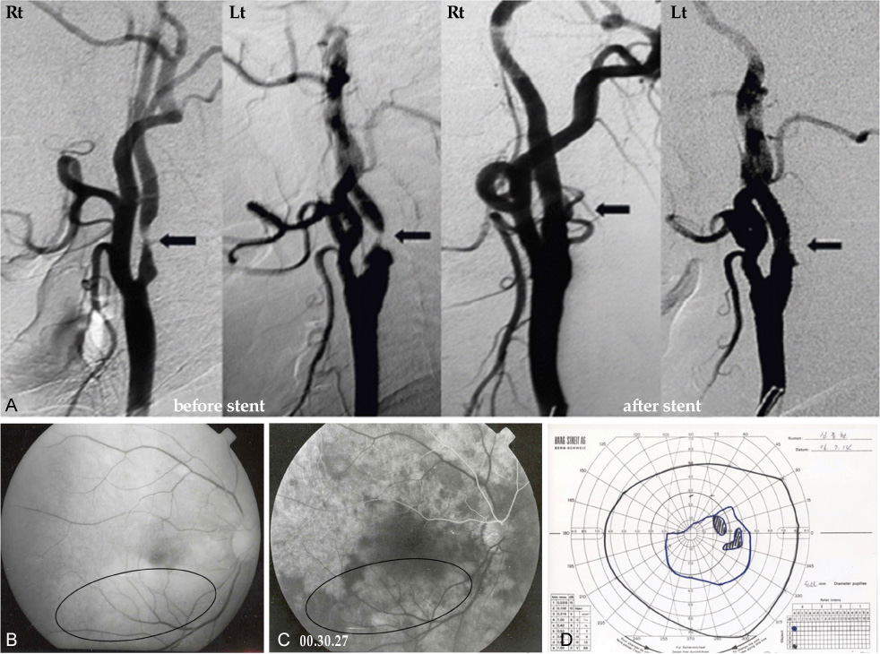
Fig. 1 (A) Trans-femoral carotid angiography shows bilateral partial occlusion of the internal carotid artery at the proximal branching portion (arrow). (B) The red-free photograph of fundus shows ischemic retina whitening at the inferior posterior pole except the fovea. (C) The fluorescein angiogram at 30 seconds after dye injection shows choroidal hypoperfusion and no filling of the retinal artery or vein in inferotemporal posterior pole. (D) The visual field test shows visual defects of the superior nasal quadrant and the superior temporal field in the right eye.
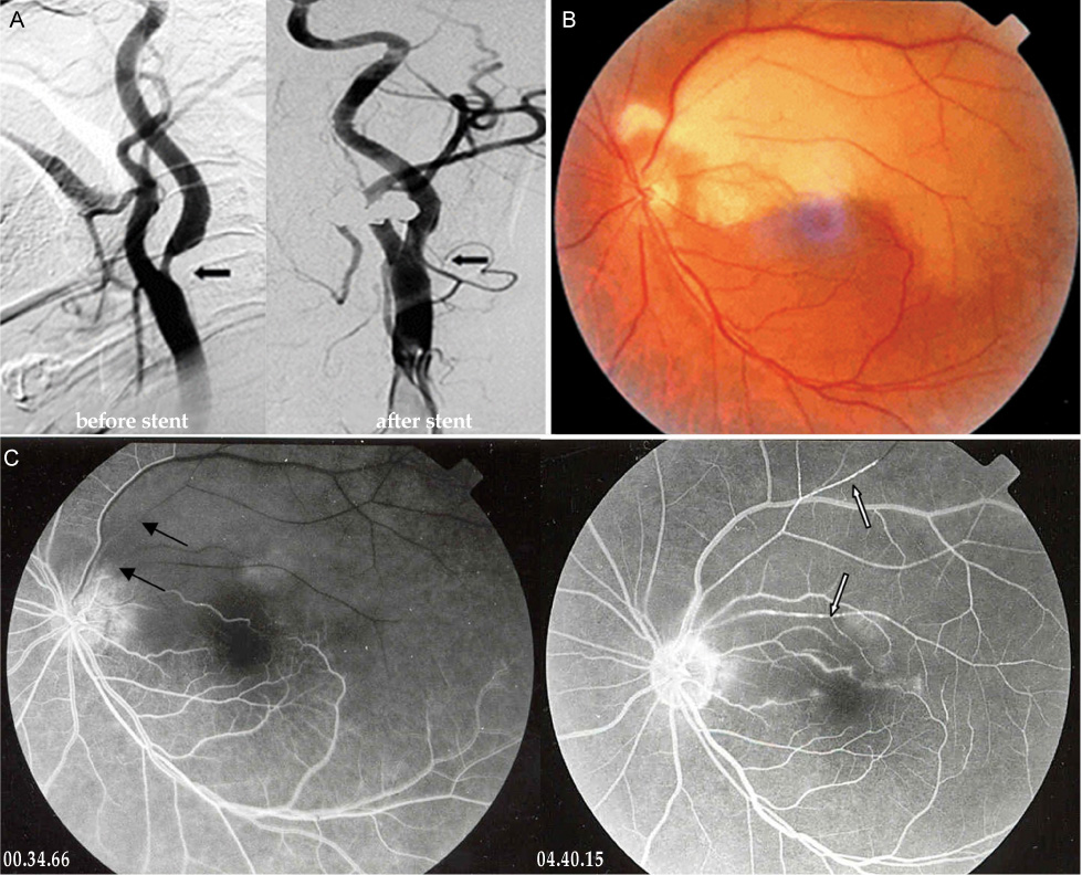
Fig. 2 (A) Trans-femoral carotid angiography shows partial occlusion of the left internal carotid artery at the proximal branching portion (arrow). (B) The color photograph of fundus shows ischemic retina whitening at the superior posterior pole except the fovea. (C) The fluorecein angiogram shows delayed filling (black arrows), embol (white arrows) and perivascular leaking in the superior temporal artery of the left eye.
Cited by 2 articles
-
Treatment of Acute Central Retinal Artery Occlusion with Ocular Ischemic Syndrome
Jong Hwan Lee, Ho Seok Moon, Dong Heun Nam, Dae Yeong Lee
J Korean Ophthalmol Soc. 2014;55(8):1242-1247. doi: 10.3341/jkos.2014.55.8.1242.Ophthalmic Artery Occlusion After Carotid Revascularization
Yeon Jin Yi, Ji Kwang Yun, Dae Won Kim, Sung Don Kang
J Cerebrovasc Endovasc Neurosurg. 2013;15(4):326-329. doi: 10.7461/jcen.2013.15.4.326.
Reference
-
1. Kimura K, Hashimoto Y, Ohno H, et al. Carotid artery Disease in Patients with Retinal Artery Occlusion. Intern med. 1996. 35:937–940.2. Wilrentz JR, Chati Z, Krafft V, Amor M. Retinal Embolization During Carotid Angioplasty and Stenting: Mechanisms and Role of Cerebral Protection Systems. Catheter Cardiovasc Interv. 2002. 56:320–327.3. Karjalainen K. Occlusion of the central retinal artery and retinal branch arterioles. A clinical, tonographic and fluorescein angiographic study of 175 patients. Acta Ophthalmol suppl. 1971. 109:1–95.4. Oshinskie L. Branch Retinal Artery Occlusion and Carotid Artery Stenosis. Am J Optom Physiol Opt. 1987. 64:144–149.5. Mark AR, Larry EM, Martin U. Branch retinal artery obstruction: a review of 201 eyes. Ann Ophthalmol. 1989. 21:103–107.6. Kim IT, Park SK, Shim SD. Continued lodging of retinal emboli in a patient with internal carotid artery and opthalmic artery occlusions. Korean J Ophthalmol. 1999. 13:36–42.7. Shin KH, Ko YI, Ko CJ. Treatment of Retinal Artery Branch Occlusion. J Korean Ophthalmol Soc. 1982. 23:209–211.8. Kim YT, Yoon IH, Won IG. A clinical study of 35 cases of retinal artery occlusion. J Korean Ophthalmol Soc. 1989. 30:119–128.9. Shimizu T, Kiyosawa M, Miura T, et al. Acute obstruction of the retinal and choroidal circulation as a complication of interventional angiography. Graefes Arch Clin Exp Ophthalmol. 1993. 231:43–47.10. Jordan WD Jr, Voellinger DC, Doblar DD, et al. Microemboli detected by transcranial Doppler monitoring in patients during carotid angioplasty versus carotid endarterectomy. Cardiovasc Surg. 1999. 7:33–38.
- Full Text Links
-
- Actions
-
Cited
- CITED
-
- Close
- Share
-
- Similar articles
-
- Central Retinal Artery Occlusion After Carotid Artery Angioplasty and Stenting in an Elderly Patient: A Case Report
- Retinal Artery Occlusion after Carotid Angioplasty and Stenting: A Case Report
- A Case of Transseptal Approach to Carotid Artery Stenting in Right Internal Carotid Stenosis
- Carotid Artery Stenting in Patients with Critical Stenosis of Proximal Internal Carotid Artery and Large Distal Arterial Thrombus: 2 Case Reports
- Percutaneous Transluminal Angioplasty with Palmaz-Schatz Stent in the Carotid Artery Stenosis
