Clear Cell "Sugar" Tumor of the Lung: A Well-Enhanced Mass with an Early Washout Pattern on Dynamic Contrast-Enhanced Computed Tomography
- Affiliations
-
- 1Department of Internal Medicine and Airway Remodeling Laboratory, Chonbuk National University Medical School, Jeonju, Korea. leeyc@chonbuk.ac.kr
- 2Department of Pathology, Chonbuk National University Medical School, Jeonju, Korea.
- 3Department of Diagnostic Radiology, Chonbuk National University Medical School, Jeonju, Korea.
- KMID: 1107544
- DOI: http://doi.org/10.3346/jkms.2008.23.6.1121
Abstract
- Clear cell tumor of the lung is a rare and very unusual benign pulmonary tumor. As clear cell tumor of the lung contains abundant cytoplasmic glycogen, this tumor is called "sugar tumor". We report a case of sugar tumor in a 64-yr-old man presenting as a round pulmonary nodule. On dynamic computed tomography (CT) scans, the solitary pulmonary nodule showed early wash-in enhancement with an early washout pattern like a lung malignancy. The patient underwent wedge resection for the tumor. Pathologic examination, including immunohistochemical studies, revealed that the nodule was a benign clear cell tumor, so-called "sugar tumor". Because only a small number of cases have been reported previously, clinical aspects, radiological characteristics on dynamic contrast-enhanced CT, and differential diagnosis of the tumor are not well established. Herein we present a clear cell tumor of the lung and discuss the clinical, radiological, and pathological features of the tumor.
MeSH Terms
-
Antigens, Neoplasm/metabolism
Diagnosis, Differential
Humans
Lung/radiography/surgery
Lung Neoplasms/diagnosis/pathology/*radiography
Male
Middle Aged
Neoplasm Proteins/metabolism
Perivascular Epithelioid Cell Neoplasms/diagnosis/pathology/*radiography
Solitary Pulmonary Nodule/diagnosis/pathology/*radiography
*Tomography, X-Ray Computed
Figure
-
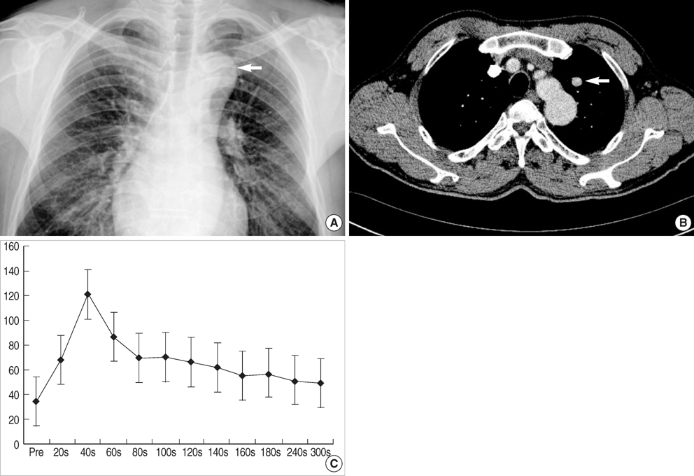
Fig. 1 Chest radiography (A) revealed an SPN in the left upper lobe. The dynamic contrast-enhanced chest CT (B) showed a well-enhanced SPN measuring 12×11×11 mm on anterior segment of left upper lobe in early phase with an early washout enhancement pattern (C). Each arrow in panel A and B indicates SPN.

Fig. 2 Macroscopic finding of the tumor. The tumor sized 12×10 mm was resected and easily enucleated. The tumor was nonencapsulated, well circumscribed, and grayish-white on cut surface.
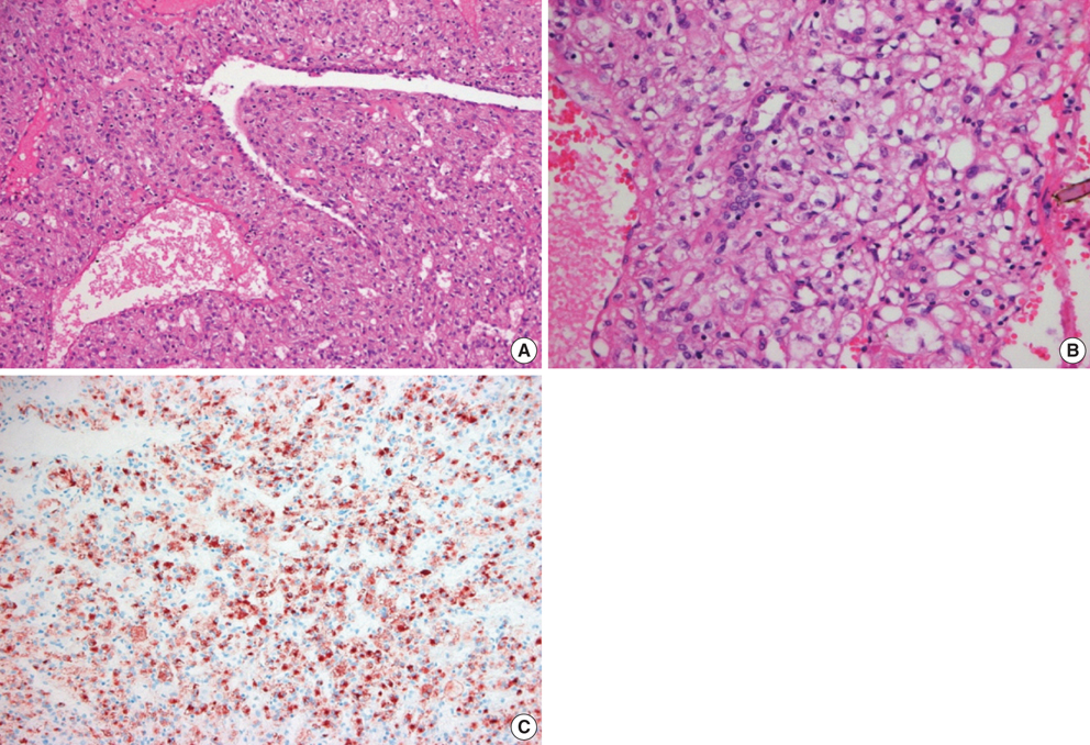
Fig. 3 Representative hematoxylin and eosin stained sections of the lung mass at low magnification (A) and at high magnification (B). The tumor cells arranged in sheets or trabuculae separated by various sized thin-walled blood vessels. In addition, the tumor cells were polygonal with abundant clear to eosinophilic cytoplasm and distinct cytoplasmic membranes. In the immunohistochemical studies, the tumor cells showed strong immunoreactivity for HMB-45 (C).
Cited by 1 articles
-
Sclerosing Perivascular Epithelioid Cell Tumor of the Lung: A Case Report with Cytologic Findings
Ha Yeon Kim, Jin Hyuk Choi, Hye Seung Lee, Yoo Jin Choi, Aeree Kim, Han Kyeom Kim
J Pathol Transl Med. 2016;50(3):238-242. doi: 10.4132/jptm.2016.02.19.
Reference
-
1. Liebow AA, Castleman B. Benign "clear cell tumors" of the lung. Am J Pathol. 1963. 43:13–14.2. Liebow AA, Castleman B. Benign clear cell ("sugar") tumors of the lung. Yale J Biol Med. 1971. 43:213–222.3. Shimosato Y, Miller RR. Biopsy interpretation of the lung. 1995. New York: Raven Press.4. Andrion A, Mazzucco G, Gugliotta P, Monga G. Benign clear cell (sugar) tumor of the lung: a light microscopic, histochemical, and ultrastructural study with a review of the literature. Cancer. 1985. 56:2657–2663.
Article5. Ozdemir IA, Zaman NU, Rullis I, Webb WR. Benign clear cell tumor of lung. J Thorac Cardiovasc Surg. 1974. 68:131–133.6. Papla B, Demczuk S, Malinowski E. Benign clear-cell "sugar" tumor of the lung-a case report. Pol J Pathol. 2003. 54:183–185.7. Gaffey MJ, Mills SE, Zarbo RJ, Weiss LM, Gown AM. Clear cell tumor of the lung. Immunohistochemical and ultrastructural evidence of melanogenesis. Am J Surg Pathol. 1991. 15:644–653.
Article8. Dail DH. Dail DH, Hammar SP, Colby TV, editors. Uncommon tumors. Pulmonary Pathology Tumors. 1995. New York: Springer;219–224.
Article9. Santana AN, Nunes FS, Ho N, Takagaki TY. A rare cause of hemoptysis: benign sugar (clear) cell tumor of the lung. Eur J Cardiothorac Surg. 2004. 25:652–654.
Article10. Gal AA, Koss MN, Hochholzer L, Chejfec G. An immunohistochemical study of benign clear cell ("sugar") tumor of the lung. Arch Pathol Lab Med. 1991. 115:1034–1038.11. Seo JB, Im JG, Seo JW, Yeon KM. Clear cell tumor of the lung. AJR Am J Roentgenol. 1996. 166:730–731.
Article12. Terra-Filho M, Kavakama J, Bagatin E, Capelozzi VL, Nery LE, Tavares R. Identification of rounded atelectasis in workers exposed to asbestos by contrast helical computed tomography. Braz J Med Biol Res. 2003. 36:1341–1347.
Article13. Swensen SJ, Brown LR, Colby TV, Weaver AL, Midthun DE. Lung nodule enhancement at CT: prospective findings. Radiology. 1996. 201:447–455.
Article14. Yi CA, Lee KS, Kim EA, Han J, Kim H, Kwon OJ, Jeong YJ, Kim S. Solitary pulmonary nodules: dynamic enhanced multi-detector row CT study and comparison with vascular endothelial growth factor and microvessel density. Radiology. 2004. 233:191–199.
Article15. Zhang M, Kono M. Solitary pulmonary nodules: evaluation of blood flow patterns with dynamic CT. Radiology. 1997. 205:471–478.
Article16. Jeong YJ, Lee KS, Jeong SY, Chung MJ, Shim SS, Kim H, Kwon OJ, Kim S. Solitary pulmonary nodule: characterization with combined wash-in and washout features at dynamic multi-detector row CT. Radiology. 2005. 237:675–683.
Article17. Jordá Aragón C, Froufe Sánchez A, Padilla Alarcón J. Benign clear cell tumor of the lung. Arch Bronconeumol. 2005. 41:59.
Article18. Gaffey MJ, Mills SE, Askin FB, Ross GW, Sale GE, Kulander BG, Visscher DW, Yousem SA, Colby TV. Clear cell tumor of the lung. A clinicopathologic, immunohistochemical, and ultrastructural study of eight cases. Am J Surg Pathol. 1990. 14:248–259.
Article19. Gaffey MJ, Mills SE, Ritter JH. Clear cell tumors of the lower respiratory tract. Semin Diagn Pathol. 1997. 14:222–232.20. Gaffey MJ, Mills SE, Frierson HF Jr, Askin FB, Maygarden SJ. Pulmonary clear cell carcinoid tumor: another entity in the differential diagnosis of pulmonary clear cell neoplasia. Am J Surg Pathol. 1998. 22:1020–1025.21. Tazelaar HD, Batts KP, Srigley JR. Primary extrapulmonary sugar tumor (PEST): a report of four cases. Mod Pathol. 2001. 14:615–622.
Article
- Full Text Links
-
- Actions
-
Cited
- CITED
-
- Close
- Share
-
- Similar articles
-
- Primary Lung Cancer: Utility of Contrast-enhanced Dynamic CT in Diagnosis with Histopathologic Correlation
- Malignant Solitary Pulmonary Nodule: Enhancement Patterns on Contrast-enhanced Dynamic CT with the Histopathologic Evaluation
- Dynamic Computed Tomography Findings of Atypical Pulmonary Hamartoma and It's Pathologic Correlations: A Case Report
- Preparative fasting before contrast-enhanced computed tomography
- Dynamic Contrast-Enhanced CT in Advanced Lung Cancer after Chemotherapy with/without Radiation Therapy: Can It Predict Treatment Responsiveness of the Tumor?
