J Korean Med Sci.
2008 Dec;23(6):1020-1026. 10.3346/jkms.2008.23.6.1020.
The Significance of Hemosiderin Deposition in the Lungs and Organs of the Mononucleated Macrophage Resorption System in Infants and Children
- Affiliations
-
- 1Uludag University Medical Faculty, Forensic Medicine Department, Bursa, Turkey. bulenteren2000@yahoo.com
- 2Council of Forensic Medicine of Turkey, Bursa, Turkey.
- 3Uludag University Medical Faculty, Department of Biostatistics, Bursa, Turkey.
- KMID: 1107528
- DOI: http://doi.org/10.3346/jkms.2008.23.6.1020
Abstract
- Hemosiderin deposition is not often recognized on routine examination with hematoxylin and eosin staining; however, iron stains may be helpful in the evaluation of hemosiderin deposition in infant autopsies. This report describes the data obtained from autopsy of 86 infants and children whose deaths were investigated at the Forensic Medicine Council Bursa Morgue Department from January 2000 to January 2003. A histochemical technique was used to identify hemosiderin in lung, liver and spleen specimens, which was correlated with other descriptive variables such as the reported cause of death, postmortem interval, trauma history, gender, and age. There was a weakly positive but significant correlation between lung and liver hemosiderin scores (Spearman's rank correlation coefficient, rho=0.348, p=0.001); i.e., given an increase in lung hemosiderin scores, an increase in liver hemosiderin scores was also observed. Similarly, a marked positive correlation between spleen and liver hemosiderin scores (Spearman's rank correlation coefficient, rho=0.335, p=0.002) was observed. The probability of spleen hemosiderin-positive cases belonging to the age group under 6 months was found to be 4.3 times greater than those who were hemosiderin-negative (95% confidence interval, 1.6-11.8). After the major differential diagnoses were ruled out, this study demonstrated, that depending on the statistically assessed morphometric grounds, the presence of hemosiderin deposits in the liver and spleen were significantly higher in the age group under 6 months.
Keyword
MeSH Terms
Figure
-
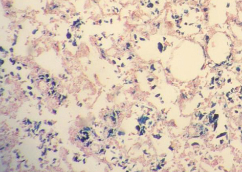
Fig. 1 Lung iron staining was predominantly found within macrophages. Perls's stain, 40× magnification.
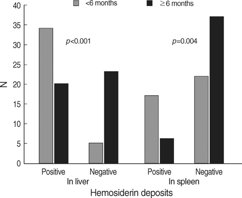
Fig. 2 Liver and spleen hemosiderin deposits in the under 6 months and older than 6 months of age groups.
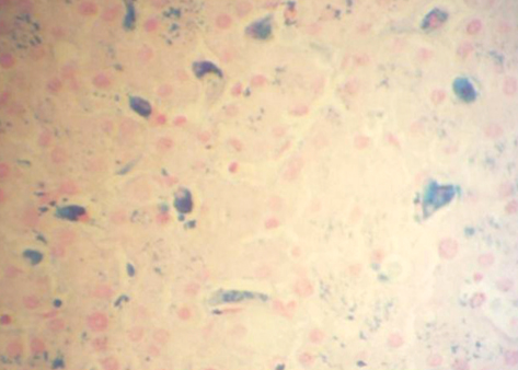
Fig. 3 Hemosiderin deposits in hepatocytes and Kupffer cells. Perls's stain, 200× magnification.
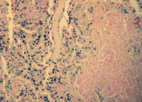
Fig. 4 Hemosiderin deposits in the spleen red pulp. Perls's stain, 200× magnification.
Reference
-
1. Hanzlick R, Delaney K. Pulmonary hemosiderin in deceased infants: baseline data for further study of infant mortality. Am J Forensic Med Pathol. 2000. 21:319–322.2. Schluckebier DA, Cool CD, Henry TE, Martin A, Wahe JW. Pulmonary siderophages and unexpected infant death. Am J Forensic Med Pathol. 2002. 23:360–363.
Article3. Betz P, Eisenmenger W. Morphometrical analysis of hemosiderin deposits in relation to wound age. Int J Legal Med. 1996. 108:262–264.
Article4. Dorandeu A, Perie G, Jouan H, Leroy B, Gray F, Durigon M. Histological demonstration of haemosiderin deposits in lungs and liver from victims of chronic physical child abuse. Int J Legal Med. 1999. 112:280–286.
Article5. Silver MM, Perrin D, Smith CR, Freedom RM. Tissue iron storage patterns in fetal hydrops associated with congestive heart failure. Pediatr Pathol Lab Med. 1996. 16:563–582.
Article6. Stewart S, Fawcett J, Jacobson W. Interstitial haemosiderin in the lungs of sudden infant death syndrome: a histological hallmark of 'ear-miss' episodes? J Pathol. 1985. 145:53–58.7. Becroft DM, Lockett BK. Intra-alveolar pulmonary siderophages in sudden infant death: a marker for previous imposed suffocation. Pathology. 1997. 29:60–63.
Article8. Milroy CM. Munchausen syndrome by proxy and intra-alveolar haemosiderin. Int J Legal Med. 1999. 112:309–312.
Article9. Centers for Disease Control and Prevention (CDC). Update: pulmonary hemorrhage/hemosiderosis among infants-Cleveland, 1993-1996. MMWR Morb Mortal Wkly Rep. 1997. 46:33–35.10. Dearborn DG. Pulmonary hemorrhage in infants and children. Curr Opin Pediatr. 1997. 9:219–224.
Article11. Becroft DM, Thompson JM, Mitchell EA. Pulmonary interstitial hemosiderin in infancy: a common consequence of normal labor. Pediatr Dev Pathol. 2005. 8:448–452.
Article12. Risse M, Weiler G. Hemosiderin findings in the liver, spleen and lung in newborn infants and infants. Z Rechtsmed. 1987. 98:181–190.13. Byard RW, Stewart WA, Telfer S, Beal SM. Assessment of pulmonary and intrathymic hemosiderin deposition in sudden infant death syndrome. Pediatr Pathol Lab Med. 1997. 17:275–282.
Article14. Lewis MJ, McKeever PK, Rutty GN. Patent ductus arteriosus as a natural cause of pulmonary hemorrhage in infants: a medicolegal dilemma. Am J Forensic Med Pathol. 2004. 25:200–204.15. Blisard KS, Bartow SA. Neonatal hemochromatosis. Hum Pathol. 1986. 17:376–383.
Article16. Sherman JM, Winnie G, Thomassen MJ, Abdul-Karim FW, Boat TF. Time course of hemosiderin production and clearance by human pulmonary macrophages. Chest. 1984. 86:409–411.
Article17. Katsumata Y, Sato K, Yada S. Green pigments in epidermal blisters of decomposed cadavers. Forensic Sci Int. 1985. 28:167–174.
Article
- Full Text Links
-
- Actions
-
Cited
- CITED
-
- Close
- Share
-
- Similar articles
-
- Forensic Pathology of the Lung in Fetus and Infant
- A Case of Hemosiderosis due to Multiple Transfusion
- Superficial Siderosis of the Central Nervous System Originating from the Thoracic Spine: A Case Report
- Transfusional Iron Overload and Choroid Plexus Hemosiderosis in a Pediatric Patient: Brain Magnetic Resonance Imaging Findings
- Lobar Intracerebral Hemorrhage Associated With Cortical Superficial Siderosis
