Pyridoxine induced neuropathy by subcutaneous administration in dogs
- Affiliations
-
- 1Department of Veterinary Internal Medicine, College of Veterinary Medicine, Seoul National University, Seoul 151-742, Korea.
- 2Department of Anatomy and Cell Biology, College of Veterinary Medicine, Seoul National University, Seoul 151-742, Korea. hyyoun@snu.ac.kr
- KMID: 1106227
- DOI: http://doi.org/10.4142/jvs.2008.9.2.127
Abstract
- To construct a sensory neuropathy model, excess pyridoxine (150 mg/kg s.i.d.) was injected subcutaneously in dogs over a period of 7 days. During the administrations period, the dogs experienced body weight reduction and proprioceptive loss involving the hindquarters. After pyridoxine administration was completed, electrophysiological recordings showed that the M wave remained at a normal state, but the H-reflex of the treated dogs disappeared at 7 days. The dorsal funiculus of L4 was disrupted irregularly in the axons and myelin with vacuolation. The dorsal root ganglia of L4, and sciatic and tibial nerves showed degenerative changes and vacuolation. However, the lateral and ventral funiculi of L4 showed a normal histopathologic pattern. Although this subcutaneous administration method did not cause systemic toxicity and effectively induced sensory neuropathy, this study confirmed the possibility of producing a pyridoxine-induced sensory neuropathy model in dogs with short-term administration.
Keyword
MeSH Terms
Figure
-
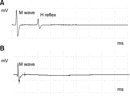
Fig. 1 On electrophysiological analysis, a control dog showed an H-reflex (A), but a pyridoxine model dog showed no H-reflex (B). Both have normal M waves. The M wave amplitude showed no remarkable change after pyridoxine treatment, a finding which was confirmed in our statistical analysis (p < 0.05). There was a remarkable change in the H-reflex after treatment in the experimental group.
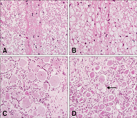
Fig. 2 (A) Normal dorsal funiculus in L4. (B) Dorsal funiculus in L4 showing disruption and irregularity of axons and myelin with vacuolation. (C) Normal dorsal root ganglia in L4. (D) Dorsal root ganglia in L4 showing chromatolysis with eccentric location of nucleus (arrow). H&E stain, ×200.

Fig. 3 (A) Normal sciatic nerve. (B) Sciatic nerve showing axonal swelling with vacuolation (arrows). (C) Normal tibial nerve. (D) Tibial nerve showing axonal swelling with vacuolation (arrows). H&E stain, ×400.
Cited by 1 articles
-
In vitro and in vivo gene therapy with CMV vector-mediated presumed dog β-nerve growth factor in pyridoxine-induced neuropathy dogs
Jin-Young Chung, Jung-Hoon Choi, Il-Seob Shin, Eun-Wha Choi, Cheol-Yong Hwang, Sang-Koo Lee, Hwa-Young Youn
J Vet Sci. 2008;9(4):367-373. doi: 10.4142/jvs.2008.9.4.367.
Reference
-
1. Callizot N, Warter JM, Poindron P. Pyridoxine-induced neuropathy in rats: a sensory neuropathy that responds to 4-methylcatechol. Neurobiol Dis. 2001. 8:626–635.
Article2. Chaudhry V, Rowinsky EK, Sartorius SE, Donehower RC, Cornblath DR. Peripheral neuropathy from taxol and cisplatin combination chemotherapy: clinical and electrophysiological studies. Ann Neurol. 1994. 35:304–311.
Article3. Dalton K, Dalton MJ. Characteristics of pyridoxine overdose neuropathy syndrome. Acta Neurol Scand. 1987. 76:8–11.
Article4. Hanrahan JP, Gordon MA. Mushroom poisoning. Case reports and a review of therapy. JAMA. 1984. 251:1057–1061.
Article5. Hoover DM, Carlton WW. The subacute neurotoxicity of excess pyridoxine HCl and clioquinol (5-chloro-7-iodo-8-hydroxyquinoline) in beagle dogs. I. Clinical disease. Vet Pathol. 1981. 18:745–756.
Article6. Hoover DM, Carlton WW. The subacute neurotoxicity of excess pyridoxine HCl and clioquinol (5-chloro-7-iodo-8-hydroxyquinoline) in beagle dogs. II. Pathology. Vet Pathol. 1981. 18:757–768.
Article7. Hopkins AP, Gilliatt RW. Motor and sensory nerve conduction velocity in the baboon: normal values and changes during acrylamide neuropathy. J Neurol Neurosurg Psychiatry. 1971. 34:415–426.
Article8. Krinke GJ, Fitzgerald RE. The pattern of pyridoxine-induced lesion: difference between the high and the low toxic level. Toxicology. 1988. 49:171–178.
Article9. Krinke G, Naylor DC, Skorpil V. Pyridoxine megavitaminosis: an analysis of the early changes induced with massive doses of vitamin B6 in rat primary sensory neurons. J Neuropathol Exp Neurol. 1985. 44:117–129.10. Perry TA, Weerasuriya A, Mouton PR, Holloway HW, Greig NH. Pyridoxine-induced toxicity in rats: a stereological quantification of the sensory neuropathy. Exp Neurol. 2004. 190:133–144.
Article11. Schaumburg H, Kaplan J, Windebank A, Vick N, Rasmus S, Pleasure D, Brown MJ. Sensory neuropathy from pyridoxine abuse. A new megavitamin syndrome. N Engl J Med. 1983. 309:445–448.12. Sims MH, Selcer RR. Occurrence and evaluation of a reflex-evoked muscle potential (H- reflex) in the normal dog. Am J Vet Res. 1981. 42:975–983.13. Xu Y, Sladky JT, Brown MJ. Dose-dependent expression of neuronopathy after experimental pyridoxine intoxication. Neurology. 1989. 39:1077–1083.
Article
- Full Text Links
-
- Actions
-
Cited
- CITED
-
- Close
- Share
-
- Similar articles
-
- Case series of pyridoxine-induced neuropathy
- Sensory Polyneuropathy Associated with Pyridoxine Overdose
- In vitro and in vivo gene therapy with CMV vector-mediated presumed dog beta-nerve growth factor in pyridoxine-induced neuropathy dogs
- Two Cases of External Ophthalmoplegia after Vincristine Treatment in Childhood
- Anaphylactic reaction after subcutaneous vitamin K1 injection in dogs: an experimental study and case report
