In vivo alternative testing with zebrafish in ecotoxicology
- Affiliations
-
- 1Department of Laboratory Animal Medicine, College of Veterinary Medicine, Seoul National University, Seoul 151-742, Korea. pjhak@snu.ac.kr
- 2Department of KRF Zoonotic Disease Priority Research Institute, College of Veterinary Medicine, Seoul National University, Seoul 151-742, Korea.
- 3Institute for Experimental Animals, College of Medicine, Seoul National University, Seoul 110-799, Korea.
- KMID: 1104909
- DOI: http://doi.org/10.4142/jvs.2008.9.4.351
Abstract
- Although rodents have previously been used in ecotoxicological studies, they are expensive, time-consuming, and are limited by strict legal restrictions. The present study used a zebrafish (Danio rerio) model and generated data that was useful for extrapolating toxicant effects in this system to that of humans. Here we treated embryos of the naive-type as well as a transiently transfected zebrafish liver cell line carrying a plasmid (phAhREEGFP), for comparing toxicity levels with the well-known aryl hydrocarbon receptor (AhR)-binding toxicants: 3,3',4,4',5-pentachlorobiphenyl (PCB126), 2,3,7,8-tetrachlorodibenzo-p-dioxin, and 3-methylcholanthrene. These toxicants induced a concentration-dependent increase in morphological disruption, indicating toxicity at early life-stages. The transient transgenic zebrafish liver cell line was sensitive enough to these toxicants to express the CYP1A1 regulated enhanced green fluorescent protein. The findings of this study demonstrated that the zebrafish in vivo model might allow for extremely rapid and reproducible toxicological profiling of early life-stage embryo development. We have also shown that the transient transgenic zebrafish liver cell line can be used for research on AhR mechanism studies.
MeSH Terms
Figure
-

Fig. 1 PCB126, TCDD, and 3-MC induced dysmorphogenesis in developing zebrafish. Embryos were immediately exposed to 10 nM PCB126 (A), 100 nM PCB126 (B, C) or 10 nM TCDD (E), 30 nM TCDD (F, G, H) or 10 µM 3-MC (I, J) or their vehicle DMSO (0.01%) (D) for 96 hr. Pericardial edema (arrows), swollen yolk sac and trunk abnormalities (A) were characterized by PCB126 toxicity. Also, PCB126-exposed zebrafish exhibited contorted tail and other tail malformations [EWE-DT238] (B, inset represents DMSO (0.01%) exposed zebrafish) and vessel irregularity (C). TCDD caused an increased incidence of trunk abnormalities, such as spinal lordosis (E). Spinal lordosis was more severe where a high concentration of TCDD was administered to the zebrafish (F). Swollen and discontinuous yolk sac was observed with TCDD exposure (G, inset represents DMSO (0.01%) exposed zebrafish). Brain hemorrhage (1), somite irregularity (2), elongated and unlooped heart (3), pericardial edema (4), no swim bladder inflation (5), swollen yolk sac (6), and lower jaw shortening (7) (arrows) were observed with TCDD exposure (H, inset represents DMSO (0.01%) exposed zebrafish). 3-MC caused pericardial edema (arrows), and swollen yolk sac (I). Also 3-MC exposed zebrafish exhibited swollen yolk sac (1), pericardial sac edema (2), elongated and unlooped heart (3), and lower jaw shortening (4) (arrows) (J). The scale bar (1 cm) in (A); the scale bar (250 µm) in (B) applies also to (G, H); the scale bar (200 µm) in (C); the scale bar (1 cm) in (D) applies also to (E, F); the scale bar (1 cm) in (I); the scale bar (250 µm) in (J).

Fig. 2 Human AhR-regulated reporter construct. phAhRE-EGFP was constructed by fusing a portion of the 5' regulatory region of the human cytochrome P4501A1 (CYP1A1) to the cDNA sequence of jellyfish GFP.
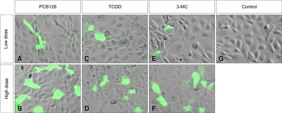
Fig. 3 EGFP expression in human AhR promoter following exposure to PCB126, TCDD, or 3-MC. ZFL cells transfected with phAhRE-EGFP and treated with 500 nM PCB126 (A) and 10 µM PCB126 (B), 20 nM TCDD (C) and 200 nM TCDD (D), 1 µM 3-MC (E) and 10 µM 3-MC (F), or their vehicle DMSO (G). Cell morphology was observed under a confocal microscope. The image merges the differential interference contrast image with the EGFP expression fluorescence image.
Reference
-
1. Ahlborg UG, Becking GC, Birnbaum LS, Brouwer A, Derks HJGM, Feeley M, Golor G, Hanberg A, Larsen JC, Liem AKD, Safe SH, Schlatter C, Waern F, Younes M, Yrjänheikki E. Toxic equivalency factors for dioxin-like PCBs. Chemosphere. 1994. 28:1049–1067.
Article2. Antkiewicz DS, Burns CG, Carney SA, Peterson RE, Heideman W. Heart malformation in an early response to TCDD in embryonic zebrafish. Toxicol Sci. 2005. 84:368–377.3. Ballschmiter K, Zell M. Analysis of polychlorinated biphenyls (PCB) by glass capillary gas chromatography. Fresenius J Anal Chem. 1980. 302:20–31.
Article4. Blechinger SR, Warren JT Jr, Kuwada JY, Krone PH. Developmental toxicology of cadmium in living embryos of a stable transgenic zebrafish line. Environ Health Perspect. 2002. 110:1041–1046.
Article5. Carney SA, Prasch AL, Heideman W, Peterson RE. Understanding dioxin developmental toxicity using the zebrafish model. Birth Defects Res A Clin Mol Teratol. 2006. 76:7–18.
Article6. Chen JN, Haffter P, Odenthal J, Vogelsang E, Brand M, van Eeden FJ, Furutani-Seiki M, Granato M, Hammerschmidt M, Heisenberg CP, Jiang YJ, Kane DA, Kelsh RN, Mullins MC, Nüsslein-Volhard C. Mutations affecting the cardiovascular system and other internal organs in zebrafish. Development. 1996. 123:293–302.
Article7. Fox K, Zauke GP, Butte W. Kinetics of bioconcentration and clearance of 28 polychlorinated biphenyl congeners in zebrafish (Brachydanio rerio). Ecotoxicol Environ Saf. 1994. 28:99–109.
Article8. Frakes RA, Zeeman CQ, Mower B. Bioaccumulation of 2,3,7,8-tetrachlorodibenzo-p-dioxin (TCDD) by fish downstream of pulp and paper mills in Maine. Ecotoxicol Environ Saf. 1993. 25:244–252.
Article9. Fraysse B, Mons R, Garric J. Developmental of a zebrafish 4-day embryo-larval bioassay to assess toxicity of chemicals. Ecotoxicol Environ Saf. 2006. 63:253–267.
Article10. Ghosh C, Zhou YL, Collodi P. Derivation and characterization of a zebrafish liver cell line. Cell Biol Toxicol. 1994. 10:167–176.
Article11. Hahn ME, Karchner SI, Shapiro MA, Perera SA. Molecular evolution of two vertebrate aryl hydrocarbon (dioxin) receptors (AHR1 and AHR2) and the PAS family. Proc Natl Acad Sci USA. 1997. 94:13743–13748.
Article12. Hope B, Scatolini S, Titus E. Bioconcentration of chlorinated biphenyls in biota from the north pacific ocean. Chemosphere. 1998. 36:1247–1261.
Article13. Hill AJ, Teraoka H, Heideman W, Peterson RE. Zebrafish as a model vertebrate for investigating chemical toxicity. Toxicol Sci. 2005. 86:6–19.
Article14. Incardona JP, Collier TK, Scholz NL. Defects in cardiac function precede morphological abnormalities in fish embryos exposed to polycyclic aromatic hydrocarbons. Toxicol Appl Phramacol. 2004. 196:191–205.
Article15. Jensen S. The PCB Story. Ambio. 1972. 1:123–131.16. Kimmel CB, Ballard WW, Kimmel SR, Ullmann B, Schilling TF. Stages of embryonic development of the zebrafish. Dev Dyn. 1995. 203:253–310.
Article17. Lele Z, Krone PH. The zebrafish as a model system in developmental, toxicological and transgenic research. Biotechnol Adv. 1996. 14:57–72.
Article18. McKim JM. Rand GM, Petrocelli SR, editors. Early life stage toxicity tests. Fundamentals of Aquatic Toxicology. 1985. New York: Hemisphere Publishing;58–95.19. Melancon MJ, Lech JJ. Uptake metabolism, and elimination of 14C-labeled 1,2,4-trichlorobenzene in rainbow trout and carp. J Toxicol Environ Health A. 1980. 6:645–658.
Article20. Mosmann T. Rapid colorimetric assay for cellular growth and survival: application to proliferation and cytotoxicity assays. J Immunol Methods. 1983. 65:55–63.
Article21. Nagel R. DarT: The embryo test with the zebrafish Danio rerio-a general model in ecotoxicology and toxicology. ALTEX. 2002. 19:Suppl 1. 38–48.22. Nebert DW, Stuart GW, Solis WA, Carvan MJ 3rd. Use of reporter genes and vertebrate DNA motifs in transgenic zebrafish as sentinels for assessing aquatic pollution. Environ Health Perspect. 2002. 110:A15.
Article23. OECD. Test No. 212: Fish, short-term toxicity test on embryo and sac-fry stages. OECD Guidelines for the Testing of Chemicals. 1998. 1:1–20.24. Ostrander GK. Techniques in Aquatic Toxicology. 1996. Boca Raton: CRC Press;517–553.25. Safe S. Polychlorinated biphenyls (PCBs), dibenzo-p-dioxins (PCDDs), dibenzofurans (PCDFs), and related compounds: Environmental and mechanistic considerations which support the development of toxic equivalency factors (TEFs). Crit Rev Toxicol. 1990. 21:51–88.
Article26. Seok SH, Park DW, Park JH, Cho SA, Baek MW, Lee HY, Kim DJ, Jin BH, Ryu DY, Park JH. β-naphthoflavone caused up-regulation of AhR regulated GFP in transgenic zebrafish. Exp Anim. 2004. 53:479–483.
Article27. Spitsbergen JM, Kent ML. The state of the art of the zebarfish model for toxicology and toxicologic pathology research-advantages and current limitations. Toxicol Pathol. 2003. 31:Suppl. 62–87.
Article28. Tanguay RL, Andreasen EA, Walker MK, Peterson RE. Schecter A, Gasiewicz TA, editors. Dioxin toxicity and aryl hydrocarbon receptor signaling in fish. Dioxins and Health. 2003. New York: John Wiley & Sons;603–628.
Article29. US Environmental Protection Agency. National Primary Drinking Water Regulations EPA 811-F-95-003-C. 1995. Ohio: National Service Center for Environmental Publications;1–62.30. Westerfield M. The Zebrafish Book. 1998. Eugene: University of Oregon Press.31. Whitlock JP Jr. Induction of cytochrome P450 1A1. Annu Rev Pharmacol Toxicol. 1999. 39:103–125.32. Zeruth G, Pollenz RS. Isolation and characterization of a dioxin inducible CYP1A1 promoter/enhancer region from zebrafish (Danio rerio). Zebrafish. 2005. 2:197–210.
Article
- Full Text Links
-
- Actions
-
Cited
- CITED
-
- Close
- Share
-
- Similar articles
-
- Zebrafish and Mycobacterial infection
- Zebrafish as an Emerging Model for Dyslipidemia and Associated Diseases
- Possibility of Zebrafish as New infection model for Leprosy
- Epigenetic profiling to environmental stressors in model and non-model organisms: Ecotoxicology perspective
- Development of a Zebrafish Larvae Model for Diabetic Heart Failure With Reduced Ejection Fraction
