Evaluating a Thrombosed Azygous Vein Aneurysm Combined with Pulmonary Arterial Thromboembolism by ECG-Gated Multidetector CT: a Case Report
- Affiliations
-
- 1Department of Radiology, Soonchunhyang University, College of Medicine, Gyeonggi-do 420-767, Korea.
- 2Department of Radiology, Chosun University Hospital, Gwangju 501-717, Korea. dhk0827@naver.com
- 3Department of Internal Medicine, Soonchunhyang University, College of Medicine, Gyeonggi-do 420-767, Korea.
- 4Department of Anesthesiology and Pain Medicine, Asan Medical Center, Ulsan University College of Medicine, Seoul 138-736, Korea.
- KMID: 1101932
- DOI: http://doi.org/10.3348/kjr.2011.12.6.754
Abstract
- Azygous vein aneurysm is a rare congenital lesion that needs to be differentiated from mediastinal mass lesions. Although almost of these anomalies are asymptomatic lesions, we experienced an interesting case in which a thrombus within an azygous vein aneurysm in a 75-year-old woman caused pulmonary thromboembolism. The patient was managed by medical treatment for one month and then the thrombus within both the azygous vein aneurysm and the pulmonary arteries completely resolved.
Keyword
MeSH Terms
Figure
-
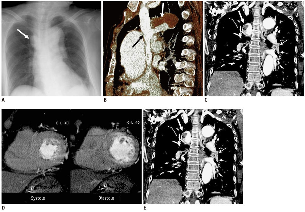
Fig. 1 75-year-old woman with azygous vein aneurysm and pulmonary thromboembolism. A. Chest plain radiograph shows well defined right paratracheal mass opacity (arrow). B. Oblique sagittal image of volume rendering reconstruction showed aneurysmal dilatation of azygous vein with thrombus (white arrow). Black arrow indicates inferior vena cava. C. Tilted coronal maximal intensity projection image of thorax shows thrombus within segmental branch of left lower pulmonary artery (small arrows) and azygous vein aneurysm (large arrow). D. Multiplanar reformatted short axis images at end-systolic phase (left image) and end-diastolic phase (right image) demonstrate normal shapes of both ventricles. Right ventricle/left ventricle diameter ratios are 0.81 at end-systolic phase and 0.75 at end-diastolic phase, respectively. E. Follow-up maximal intensity projection image after anticoagulation treatment for one month shows resolved thrombus within segmental branch of left lower pulmonary artery as compared with that of C. There is subsegmental atelectasis in left lower lung field.
Reference
-
1. Nakamura Y, Nakano K, Nakatani H, Fukuda T, Honda K, Homma N. Surgical exclusion of a thrombosed azygos vein aneurysm causing pulmonary embolism. J Thorac Cardiovasc Surg. 2007. 133:834–835.2. Ishikura H, Kimura S, Fukumura Y, Ohtani T. Resection of an azygos vein aneurysm with thrombosis. Gen Thorac Cardiovasc Surg. 2010. 58:209–211.3. Mehta M, Towers M. Computed tomography appearance of idiopathic aneurysm of the azygos vein. Can Assoc Radiol J. 1996. 47:288–290.4. Jain A, Blebea JS. Post-traumatic pseudoaneurysm of the azygous vein in a patient with azygous continuation. J Comput Assist Tomogr. 1994. 18:647–648.5. Chiu SS, Lau S, Kam CK. Azygous vein aneurysm: CT scan follow-up. J Thorac Imaging. 2006. 21:66–68.6. Podbielski FJ, Sam AD 2nd, Halldorsson AO, Iasha-Sznajder J, Vigneswaran WT. Giant azygos vein varix. Ann Thorac Surg. 1997. 63:1167–1169.7. Watanabe A, Kusajima K, Aisaka N, Sugawara H, Tsunematsu K. Idiopathic saccular azygos vein aneurysm. Ann Thorac Surg. 1998. 65:1459–1461.8. Kurihara Y, Nakajima Y, Ishikawa T. Case report: saccular aneurysm of the azygos vein simulating a paratracheal tumour. Clin Radiol. 1993. 48:427–428.9. Gnanamuthu BR, Tharion J. Azygos vein aneurysm--a case for elective resection. Heart Lung Circ. 2008. 17:62–64.10. Lu MT, Cai T, Ersoy H, Whitmore AG, Levit NA, Goldhaber SZ, et al. Comparison of ECG-gated versus non-gated CT ventricular measurements in thirty patients with acute pulmonary embolism. Int J Cardiovasc Imaging. 2009. 25:101–107.
- Full Text Links
-
- Actions
-
Cited
- CITED
-
- Close
- Share
-
- Similar articles
-
- Fatal Pulmonary Thromboembolism Caused by Popliteal Vein Aneurysm
- Pulmonary Embolism Caused by Popliteal Venous Aneurysm
- Unilateral Pulmonary Vein Atresia: A Case Report
- Cystic Lung Changes in a Thin Section CT in an Asymptomatic Young Adult with Unilateral Pulmonary Vein Atresia: A Case Report
- A Case of Pulmonary Thromboembolism in Active Ulcerative Colitis
