A Case of Ciliary Body Melanocytoma Presenting as a Painful Iris Mass
- Affiliations
-
- 1Department of Ophthalmology, Institute of Vision Research, Yonsei University College of Medicine, Seoul, Korea. sunglee@yumc.yonsei.ac.kr
- 2Department of Pathology, Yonsei University College of Medicine, Seoul, Korea.
- KMID: 1098793
- DOI: http://doi.org/10.3341/kjo.2010.24.1.44
Abstract
- We report a case of ciliary body melanocytoma in a Korean patient, which presented as an intermittently painful pigmented iris mass and was successfully managed by iridocyclectomy. A 52-year-old healthy man presented with an irregularly-shaped and heavily-pigmented mass at the iris root of his right eye. Visual acuity of the right eye was 20/20 with normal intraocular pressure. Ultrasound biomicroscopy showed a 1.5x1.3-mm ciliary-body mass with extension into the iris root. Iridocyclectomy with scleral resection under a lamellar scleral flap was performed, and the histopathologic features of the resected tissue were consistent with melanocytoma of the ciliary body. The patient's visual acuity remained 20/20 with good postoperative cosmesis. During one year of follow-up, no signs of tumor recurrence were seen, and the patient reported resolution of the intermittent ocular pain in the involved eye.
Keyword
MeSH Terms
Figure
-
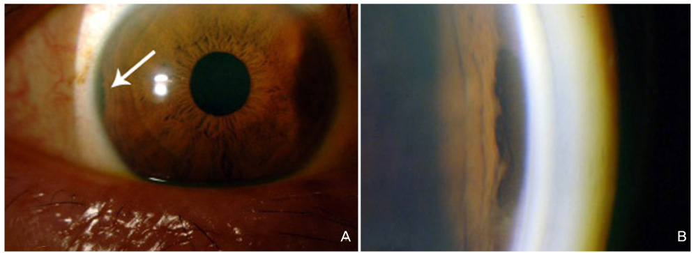
Fig. 1 (A) Colored slit-lamp photograph showing a dark-pigmented and irregularly-shaped mass at the temporal iris root (white arrow) in the right eye. (B) Gonioscopic examination showing the mass in an enlarged view.
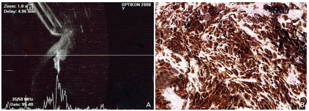
Fig. 2 (A) Ultrasound biomicroscopy shows a ciliary body mass with extension into the iris root. Medium internal reflection of the mass was seen with a basal diameter of 1.5 mm and a height of 1.3 mm. (B) Microscopic appearance of the specimen showing polyhedral cells filled with dark melanin granules, typical features of a melanocytoma.
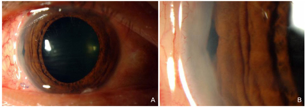
Fig. 3 Slit-lamp photograph taken on postoperative day 7. (A) Note a small opening at the iris root with good overall postoperative cosmesis. (B) Enlarged view of the iridectomy site.
Cited by 1 articles
-
A Case of Melanocytoma Originating from the Iris
Jin Ah Lee, Yong Sun Ahn, Yang Kyung Cho
J Korean Ophthalmol Soc. 2014;55(5):789-793. doi: 10.3341/jkos.2014.55.5.789.
Reference
-
1. Demirci H, Mashayekhi A, Shields CL, et al. Iris melanocytoma: clinical features and natural course in 47 cases. Am J Ophthalmol. 2005. 139:468–475.2. Brownstein S, Dorey MW, Mathew B, et al. Melanocytoma of the choroid: atypical presentation and review of the literature. Can J Ophthalmol. 2002. 37:247–252.3. Shields JA, Shields CL, Eagle RC Jr. Melanocytoma (hyperpigmented magnocellular nevus) of the uveal tract: the 34th G. Victor Simpson lecture. Retina. 2007. 27:730–739.4. LoRusso FJ, Boniuk M, Font RL. Melanocytoma (magnocellular nevus) of the ciliary body: report of 10 cases and review of the literature. Ophthalmology. 2000. 107:795–800.5. Capeans C, Pineiro A, Blanco MJ, et al. Ultrasound biomicroscopic findings in a cavitary melanocytoma of the ciliary body. Can J Ophthalmol. 2003. 38:501–503.6. Haruta M, Miyamoto K, Horii T, et al. Iridociliary melanocytoma with suspected pulmonary metastasis. Jpn J Ophthalmol. 2005. 49:545–547.7. Hiscott P, Campbell RJ, Robertson DM, Damato B. Intraocular melanocytoma in association with bone formation. Arch Ophthalmol. 2003. 121:1791–1794.8. Mohamed MD, Gupta M, Parsons A, Rennie IG. Ultrasound biomicroscopy in the management of melanocytoma of the ciliary body with extrascleral extension. Br J Ophthalmol. 2005. 89:14–16.9. Wei WB, Yang WL, Hu SM, Li B. Local excision of ciliary body tumors: a long-term prospective cohort study in China. Chin Med J. 2008. 121:2152–2156.
- Full Text Links
-
- Actions
-
Cited
- CITED
-
- Close
- Share
-
- Similar articles
-
- A Case of Melanocytoma of the Ciliary Body
- A Case of Melanocytoma Originating from the Iris
- IgG-lambda extramedullary Plasmacytoma of the Anterior Uveal Tract: Case Report
- The Morphological Change of Iris and Ciliary Body in the Korean Fetal Eyes according to Gestational Age
- A Study on Mitomycin C Induced Damage to the Iris and Ciliary Body of Cats
