Pulsed and Color Doppler Sonographic Findings of Penile Mondor's Disease
- Affiliations
-
- 1Department of Radiology, University of Konyang School of Medicine, Daejeon, Korea. bookdoo7@chollian.net
- KMID: 1098198
- DOI: http://doi.org/10.3348/kjr.2008.9.2.179
Abstract
- This report describes the color and pulsed Doppler US findings of penile Mondor's disease. The pulsed Doppler US findings of penile Mondor's disease have not been previously published, so we report here for the first time on the cavernosal arterial flow signal pattern of penile Mondor's disease. Penile Mondor's disease is rare disease that's characterized by thrombosis in the dorsal vein of the penis. The previous reports on penile Mondor's disease are concerned with the color Doppler US finding without the flow signals in this area, but these findings are insufficient to understand the hemodynamics in penile Mondor's disease. We report for the first time on a cavernosal artery flow signal pattern of low peak systolic velocity and high-resistance.
MeSH Terms
Figure
-
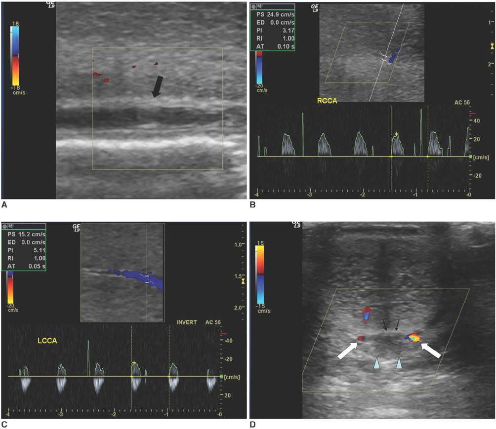
Fig. 1 Penile Mondor's disease in 38-year-old man. A. Grey scale US revealed internal echogenicity in penile superficial dorsal vein (arrow). Color Doppler US failed to detect venous flow signals in this area. B, C. Pulsed wave Doppler US without intracavernosal vasoactive agent administration showed weak flow signal with low peak systolic velocity and high-resistance pattern. Right (B) and left side (C) peak systolic velocities of carvernosal artery were 24.9 and 16 cm/sec, respectively, and resistance indexes in both sides were 1.0. D. Thirty minutes after intracavernosal vasoactive agent injection, Doppler US showed flow signal in bilateral dorsal arteries (white arrows), but no flow in either superficial dorsal (arrowheads) or deep dorsal veins (black arrows).
Reference
-
1. Mondor H. Tronculite sous-cutanée subaiguë de la paroi thoracigue antéro-latérale. Mem Acad Chir. 1939. 65:1271–1278.2. Braun-Falco O. Clinical manifestations, histology and pathogenesis of the cordlike superficial phlebitis forms. Dermatol Wochenschr. 1955. 132:705–715.3. Helm JD Jr, Hodge IG. Thrombophlebitis of a dorsal vein of the penis: report of a case treated by phenylbutazone (Butazolidin). J Urol. 1958. 79:306–307.4. Rodriguez Faba O, Parra Muntaner L, Gómez Cisneros SC, Martin Benito JL, Escaf Barmadah S. Thrombosis of the dorsal penis vein (of Mondor's phlebitis). Presentation of a new case. Actas Urol Esp. 2006. 30:80–82.5. Koh JS, Suh HJ, Choe HS, Jung JH, Kim YS, Kim JA, et al. Superficial thrombophlebitis of the dorsal vein of the penis(Penile Mondor's disease). Korean J Urol. 2004. 45:399–401.6. Al-Mwalad M, Loertzer H, Wicht A, Fornara P. Subcutaneous penile vein thrombosis (Penile Mondor's Disease): Pathogenesis, diagnosis, and therapy. Urology. 2006. 67:586–588.7. Foshager MC, Hedlund LJ, Troppmann C, Benedetti E, Gruessner RW. Venous thrombosis of pancreatic transplants: diagnosis by duplex sonography. AJR Am J Roentgenol. 1997. 169:1269–1273.8. Reuther G, Wanjura D, Bauer H. Acute renal vein thrombosis in renal allografts: detection with duplex Doppler US. Radiology. 1989. 170:557–558.9. Fitzgerald SW, Erickson SJ, Foley DW, Lipchik EO, Lawson TL. Color Doppler sonography in the evaluation of erectile dysfunction. Radiographics. 1992. 12:3–17.
- Full Text Links
-
- Actions
-
Cited
- CITED
-
- Close
- Share
-
- Similar articles
-
- Penile Emergencies– Demystifying the Sonographic Spectrum
- Thrombophlebitis of the Penile Superficial Vein, Penile Mondor's Disease: A Case Report
- Penile Doppler ultrasonography revisited
- Prolonged oral sildenafil use-induced Mondor disease: a case report
- Doppler ultrasonography of the lower extremity arteries: anatomy and scanning guidelines
